

Herein, we report two cases of 2019 novel coronavirus (2019-nCoV). Both patients are believed to have exposure related to Wuhan, China (1–4). We demonstrated change in the course of disease over time on CT scans (5,6).
Case 1
A 45-year-old woman living in Wuhan, China, was admitted to the hospital for 2 days due to cough and fever. On admission, her body temperature was elevated (38.2°C [100.76°F]) and there were coarse breath sounds of both lungs at auscultation. Her laboratory studies showed an increased neutrophil ratio (81.2%; normal range, 40.0%–75.0%), decreased lymphocyte ratio (12.8%; normal range, 20.0%–50.0%), increased erythrocyte sedimentation rate (24 mm/h; normal range, <20 mm/h), normal D-dimer concentration, and increased lymphokine interleukin 6 (27.47 pg/mL; normal range, 0.1–2.9 pg/mL). Real-time fluorescence polymerase chain reaction of the patient’s sputum was positive for the 2019-nCoV nucleic acid. Unenhanced chest CT showed multiple bilateral areas of peripheral consolidation (Fig 1a, 1b). There was interlobular septal thickening with a crazy paving appearance and bronchiectasis. The adjacent pleura was thickened, without mediastinal lymphadenopathy or pleural fluid. Two days after admission, the patient’s temperature rose to 39.2°C (102.56°F) and constitutional symptoms worsened. Repeat chest CT showed progressive consolidation with loss of the areas of crazy paving (Fig 1c, 1d). After antiviral and symptomatic treatment, the patient remains under close observation with improvement in symptoms.
Figure 1a:
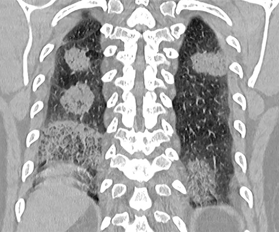
Unenhanced chest CT images in a 45-year-old woman with 2019 novel coronavirus (2019-nCoV) pneumonia. (a) Coronal CT scan shows multiple bilateral areas of consolidation with some central low attenuation suggesting an organizing pneumonia pattern. (b) Axial image shows the right lower lobe organizing pneumonia and an early crazy-paving pattern in the left lower lobe. (c) CT coronal reformation obtained 2 days later shows that the bilateral consolidations have increased in CT value. (d) Corresponding axial CT scan shows that the crazy-paving pattern has resolved.
Figure 1b:
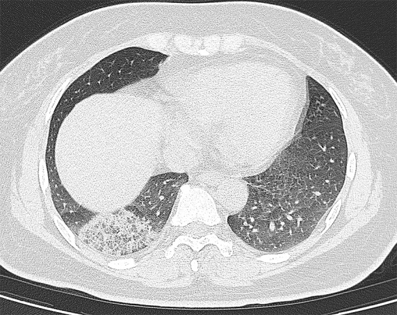
Unenhanced chest CT images in a 45-year-old woman with 2019 novel coronavirus (2019-nCoV) pneumonia. (a) Coronal CT scan shows multiple bilateral areas of consolidation with some central low attenuation suggesting an organizing pneumonia pattern. (b) Axial image shows the right lower lobe organizing pneumonia and an early crazy-paving pattern in the left lower lobe. (c) CT coronal reformation obtained 2 days later shows that the bilateral consolidations have increased in CT value. (d) Corresponding axial CT scan shows that the crazy-paving pattern has resolved.
Figure 1c:
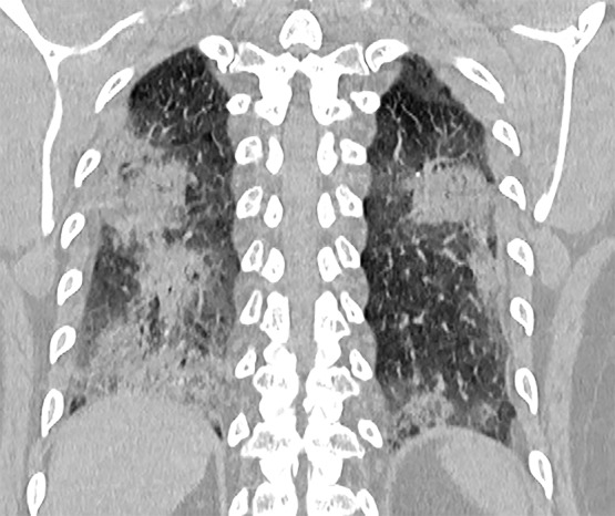
Unenhanced chest CT images in a 45-year-old woman with 2019 novel coronavirus (2019-nCoV) pneumonia. (a) Coronal CT scan shows multiple bilateral areas of consolidation with some central low attenuation suggesting an organizing pneumonia pattern. (b) Axial image shows the right lower lobe organizing pneumonia and an early crazy-paving pattern in the left lower lobe. (c) CT coronal reformation obtained 2 days later shows that the bilateral consolidations have increased in CT value. (d) Corresponding axial CT scan shows that the crazy-paving pattern has resolved.
Figure 1d:
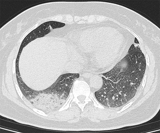
Unenhanced chest CT images in a 45-year-old woman with 2019 novel coronavirus (2019-nCoV) pneumonia. (a) Coronal CT scan shows multiple bilateral areas of consolidation with some central low attenuation suggesting an organizing pneumonia pattern. (b) Axial image shows the right lower lobe organizing pneumonia and an early crazy-paving pattern in the left lower lobe. (c) CT coronal reformation obtained 2 days later shows that the bilateral consolidations have increased in CT value. (d) Corresponding axial CT scan shows that the crazy-paving pattern has resolved.
Case 2
A 32-year-old man had a history of business travel to Wuhan, China. He was admitted to the hospital with cough and fever. At admission, his body temperature was slightly elevated (38.1°C [100.58°F]) and coarse breath sounds of both lungs were found at auscultation. Laboratory studies showed normal complete blood count values, normal C-reactive protein, and normal D-dimer concentration. The lymphokine interleukin 6 level was increased (413.6 pg/mL; normal range, 0.1–2.9 pg/mL). Real-time fluorescence polymerase chain reaction of the patient’s sputum was positive for 2019-nCoV nucleic acid. Unenhanced chest CT showed subpleural right lower lobe consolidation (Fig 2a). There was bronchiectasis with reactive thickening of the adjacent pleura. There was no mediastinal lymphadenopathy or pleural effusion. After 3 days of antiviral and symptomatic treatment, the right lower lobe consolidation increased (Fig 2b). Repeat chest CT 8 days later (Fig 2c) showed improvement. The patient recovered uneventfully after 10 days.
Figure 2a:
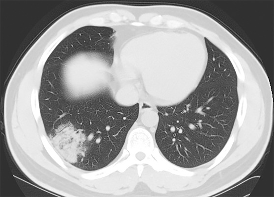
Unenhanced chest CT images in a 32-year-old man with 2019 novel coronavirus (2019-nCoV) pneumonia. (a) CT scan shows a right lower lobe subpleural consolidation. (b) CT scan obtained 3 days later shows that the right lower lobe consolidation has evolved into more of an organizing pneumonia pattern. (c) CT scan obtained after 8 days of therapy shows near-complete resolution of the right lower lobe airspace disease.
Figure 2b:
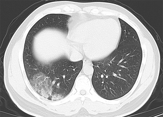
Unenhanced chest CT images in a 32-year-old man with 2019 novel coronavirus (2019-nCoV) pneumonia. (a) CT scan shows a right lower lobe subpleural consolidation. (b) CT scan obtained 3 days later shows that the right lower lobe consolidation has evolved into more of an organizing pneumonia pattern. (c) CT scan obtained after 8 days of therapy shows near-complete resolution of the right lower lobe airspace disease.
Figure 2c:
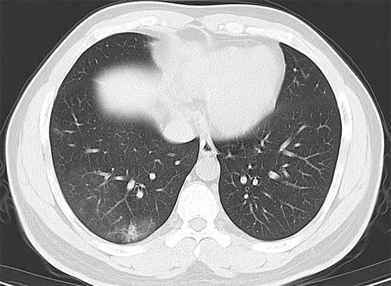
Unenhanced chest CT images in a 32-year-old man with 2019 novel coronavirus (2019-nCoV) pneumonia. (a) CT scan shows a right lower lobe subpleural consolidation. (b) CT scan obtained 3 days later shows that the right lower lobe consolidation has evolved into more of an organizing pneumonia pattern. (c) CT scan obtained after 8 days of therapy shows near-complete resolution of the right lower lobe airspace disease.
Footnotes
Disclosures of Conflicts of Interest: Y.F. disclosed no relevant relationships. H.Z. disclosed no relevant relationships. Y.X. disclosed no relevant relationships. J.X. disclosed no relevant relationships. P.P. disclosed no relevant relationships. W.J. disclosed no relevant relationships.
References
- 1.Holshue ML, DeBolt C, Lindquist S, et al. First Case of 2019 Novel Coronavirus in the United States. N Engl J Med doi:10.1056/NEJMoa2001191. Published online January 31, 2020. [DOI] [PMC free article] [PubMed]
- 2.Li Q, Guan X, Wu P, Wang X, et al. Early Transmission Dynamics in Wuhan, China, of Novel Coronavirus-Infected Pneumonia. N Engl J Med doi:10.1056/NEJMoa2001316. Published online January 29, 2020. [DOI] [PMC free article] [PubMed]
- 3.Huang C, Wang Y, Li X, et al. Clinical features of patients infected with 2019 novel coronavirus in Wuhan, China. Lancet doi:10.1016/S0140-6736(20)30183-5. Published online January 24, 2020. [DOI] [PMC free article] [PubMed]
- 4.Lei J, Li J, Li X, Qi X. CT Imaging of the 2019 Novel Coronavirus (2019-nCoV) Pneumonia. Radiology 2020;295:18. [DOI] [PMC free article] [PubMed] [Google Scholar]
- 5.Chung M, Bernheim A, Mei X. et al. CT imaging features of 2019 novel coronavirus (2019-nCoV). Radiology: 2020;295:202–207. [DOI] [PMC free article] [PubMed] [Google Scholar]
- 6.Song F, Shi N, Shan F, et al. Emerging 2019 novel coronavirus (2019-nCoV) pneumonia. Radiology 2020;295:210–217. [DOI] [PMC free article] [PubMed] [Google Scholar]