All articles published by MDPI are made immediately available worldwide under an open access license. No special
permission is required to reuse all or part of the article published by MDPI, including figures and tables. For
articles published under an open access Creative Common CC BY license, any part of the article may be reused without
permission provided that the original article is clearly cited. For more information, please refer to
https://www.mdpi.com/openaccess.
Feature papers represent the most advanced research with significant potential for high impact in the field. A Feature
Paper should be a substantial original Article that involves several techniques or approaches, provides an outlook for
future research directions and describes possible research applications.
Feature papers are submitted upon individual invitation or recommendation by the scientific editors and must receive
positive feedback from the reviewers.
Editor’s Choice articles are based on recommendations by the scientific editors of MDPI journals from around the world.
Editors select a small number of articles recently published in the journal that they believe will be particularly
interesting to readers, or important in the respective research area. The aim is to provide a snapshot of some of the
most exciting work published in the various research areas of the journal.
Shemyakin-Ovchinnikov Institute of Bioorganic Chemistry, Russian Academy of Sciences, 117997 Moscow, Russia Interests: exosomes; tumor therapy; nanoparticles; PD1\PD-L1 therapy; mouse models
Special Issue Information
Dear Colleagues,
Mesenchymal stem cell (MSC) clinical application is hindered by the poorly identified molecular mechanisms of MSC action. An improvement was achieved with the identification of the clinical efficacy of MSC exosomes. However, neither the content of different source exosomes nor the molecular mechanisms of their action are known. Exosomes vary in size and content and carry different sets of molecules: RNA, DNA, proteins, lipids. Exosomal proteins are represented mostly by the membrane anchored proteins, while multiple hydrophilic MSC proteins can also be of use. The aim of this Special Issue is to collect results on MSC and studies of their byproducts and clinical applications focused on the possible mechanisms of their action.
Dr. Elena V. Svirshchevskaya Guest Editor
Manuscript Submission Information
Manuscripts should be submitted online at www.mdpi.com by registering and logging in to this website. Once you are registered, click here to go to the submission form. Manuscripts can be submitted until the deadline. All submissions that pass pre-check are peer-reviewed. Accepted papers will be published continuously in the journal (as soon as accepted) and will be listed together on the special issue website. Research articles, review articles as well as short communications are invited. For planned papers, a title and short abstract (about 100 words) can be sent to the Editorial Office for announcement on this website.
Submitted manuscripts should not have been published previously, nor be under consideration for publication elsewhere (except conference proceedings papers). All manuscripts are thoroughly refereed through a single-blind peer-review process. A guide for authors and other relevant information for submission of manuscripts is available on the Instructions for Authors page. Current Issues in Molecular Biology is an international peer-reviewed open access monthly journal published by MDPI.
by
Daria S. Chulpanova, Tamara V. Pukhalskaia, Zarema E. Gilazieva, Yuliya V. Filina, Milana N. Mansurova, Albert A. Rizvanov and Valeriya V. Solovyeva
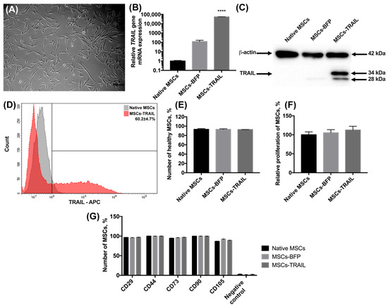
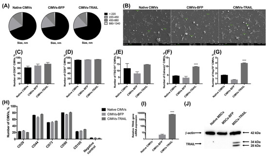
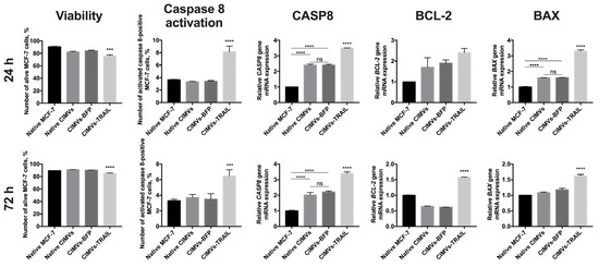
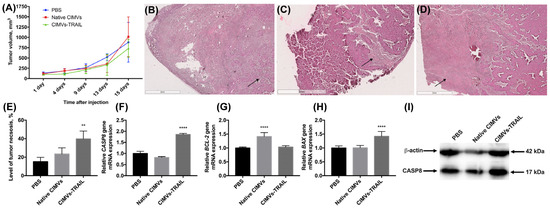
Tumor-necrosis-factor-associated apoptosis-inducing ligand (TRAIL) is one of the most promising therapeutic cytokines that selectively induce apoptosis in tumor cells. It is known that membrane vesicles (MVs) can carry the surface markers of parental cells. Therefore, MVs are of interest as a tool for
[...] Read more.
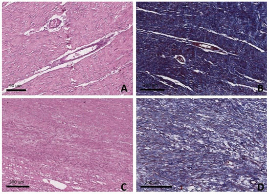
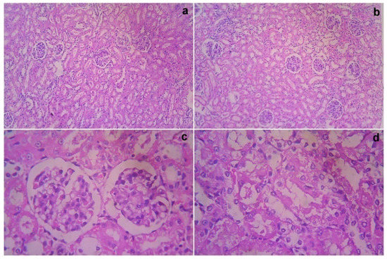
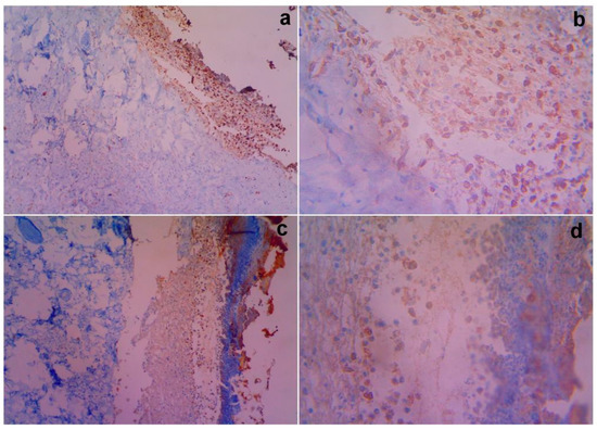
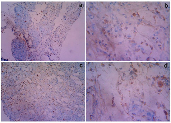
Tumor-necrosis-factor-associated apoptosis-inducing ligand (TRAIL) is one of the most promising therapeutic cytokines that selectively induce apoptosis in tumor cells. It is known that membrane vesicles (MVs) can carry the surface markers of parental cells. Therefore, MVs are of interest as a tool for cell-free cancer therapy. In this study, membrane vesicles were isolated from TRAIL-overexpressing mesenchymal stem cells using cytochalasin B treatment (CIMVs). To evaluate the antitumor effect of CIMVs-TRAIL in vivo, a breast cancer mouse model was produced. The animals were intratumorally injected with 50 µg of native CIMVs or CIMVs-TRAIL for 12 days with an interval of two days. Then, tumor growth rate, tumor necrotic area, the expression of the apoptosis-related genes CASP8, BCL-2, and BAX and the level of CASP8 protein were analyzed. A 1.8-fold increase in the CAS8 gene mRNA and a 1.7-fold increase in the CASP8 protein level were observed in the tumors injected with CIMVs-TRAIL. The expression of the anti-apoptotic BCL-2 gene in the CIMV-TRAIL group remained unchanged, while the mRNA level of the pro-apoptotic BAX gene was increased by 1.4 times, which indicated apoptosis activation in the tumor tissue. Thus, CIMVs-TRAIL were able to activate the extrinsic apoptosis pathway and induce tumor cell death in the breast cancer mouse model.
Full article
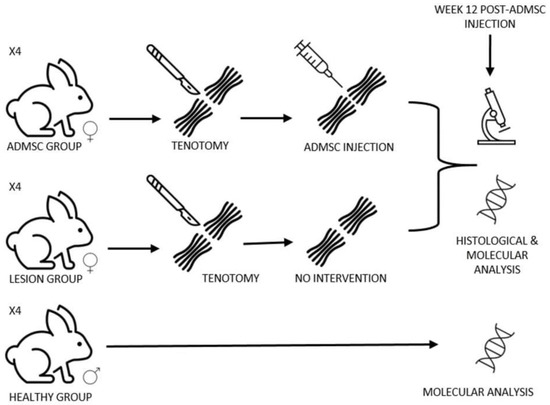
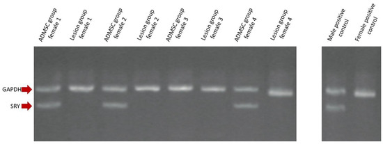
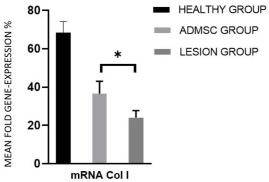
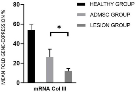
Background: Achilles-tendon rupture prevails as a common tendon pathology. Adipose-derived mesenchymal stem cells (ADMSCs) are multipotent stem cells derived from adipose tissue with attractive regeneration properties; thus, their application in tendinopathies could be beneficial. Methods: Male rabbit ADMSCs were obtained from the falciform
[...] Read more.
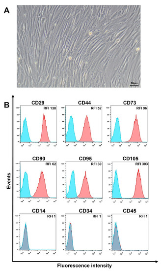
Background: Achilles-tendon rupture prevails as a common tendon pathology. Adipose-derived mesenchymal stem cells (ADMSCs) are multipotent stem cells derived from adipose tissue with attractive regeneration properties; thus, their application in tendinopathies could be beneficial. Methods: Male rabbit ADMSCs were obtained from the falciform ligament according to previously established methods. After tenotomy and suture of the Achilles tendon, 1 × 10⁶ flow-cytometry-characterized male ADMSCs were injected in four female New Zealand white rabbits in the experimental group (ADMSC group), whereas four rabbits were left untreated (lesion group). Confirmation of ADMSC presence in the injured site after 12 weeks was performed with quantitative sex-determining region Y (SRY)-gene RT-PCR. At Week 12, histochemical analysis was performed to evaluate tissue regeneration along with quantitative RT-PCR of collagen I and collagen III mRNA. Results: Presence of male ADMSCs was confirmed at Week 12. No statistically significant differences were found in the histochemical analysis; however, statistically significant differences between ADMSC and lesion group expression of collagen I and collagen III were evidenced, with 36.6% and 24.1% GAPDH-normalized mean expression, respectively, for collagen I (p < 0.05) and 26.3% and 11.9% GAPDH-normalized mean expression, respectively, for collagen III (p < 0.05). The expression ratio between the ADMSC and lesion group was 1.5 and 2.2 for collagen I and collagen III, respectively. Conclusion: Our results make an important contribution to the understanding and effect of ADMSCs in Achilles-tendon rupture.
Full article
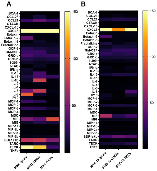
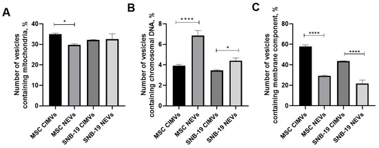
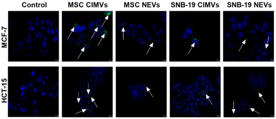
To date, there are numerous protocols for the isolation of extracellular vesicles (EVs). Depending on the isolation method, it is possible to obtain vesicles with different characteristics, enriched with specific groups of proteins, DNA and RNA, which affect similar types of cells in
[...] Read more.
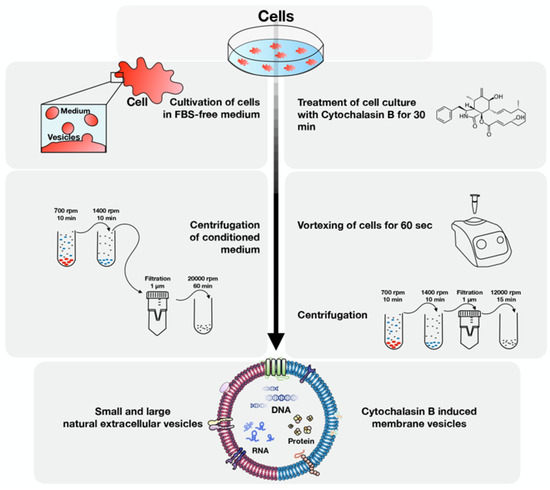
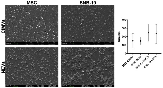
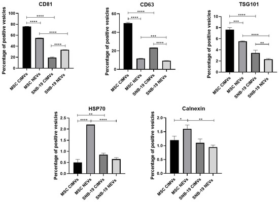
To date, there are numerous protocols for the isolation of extracellular vesicles (EVs). Depending on the isolation method, it is possible to obtain vesicles with different characteristics, enriched with specific groups of proteins, DNA and RNA, which affect similar types of cells in the opposite way. Therefore, it is important to study and compare methods of vesicle isolation. Moreover, the differences between the EVs derived from tumor and mesenchymal stem cells are still poorly understood. This article compares EVs from human glioblastoma cells and mesenchymal stem cells (MSCs) obtained by two different methods, ultracentrifugation and cytochalasin B-mediated induction. The size of the vesicles, the presence of the main EV markers, the presence of nuclear and mitochondrial components, and the molecular composition of the vesicles were determined. It has been shown that EVs obtained by both ultracentrifugation and cytochalasin B treatment have similar features, contain particles of endogenous and membrane origin and can interact with monolayer cultures of tumor cells.
Full article
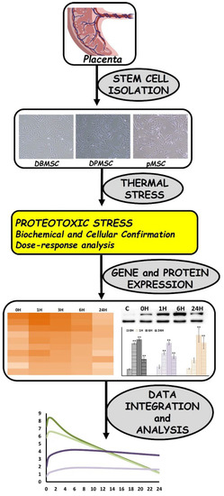
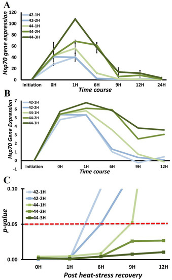
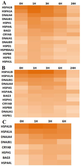
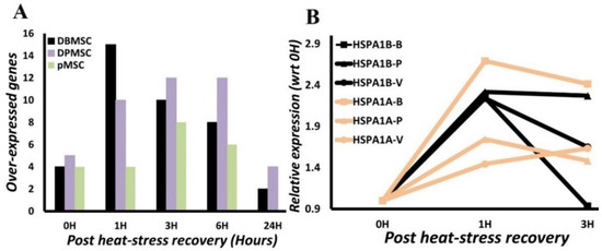
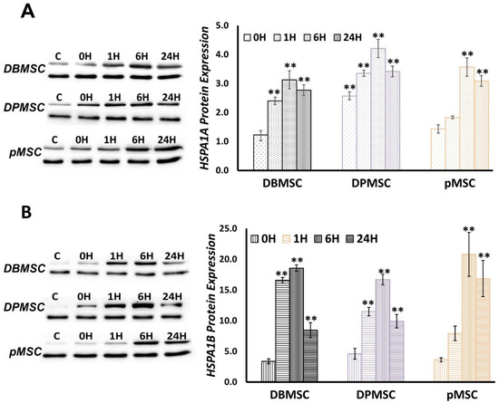
Placenta-derived stem cells (PDSCs), due to unique traits such as mesenchymal and embryonic characteristics and the absence of ethical constraints, are in a clinically and therapeutically advantageous position. To aid in stemness maintenance, counter pathophysiological stresses, and withstand post-differentiation challenges, stem cells require
[...] Read more.
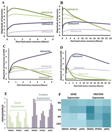
Placenta-derived stem cells (PDSCs), due to unique traits such as mesenchymal and embryonic characteristics and the absence of ethical constraints, are in a clinically and therapeutically advantageous position. To aid in stemness maintenance, counter pathophysiological stresses, and withstand post-differentiation challenges, stem cells require elevated protein synthesis and consequently augmented proteostasis. Stem cells exhibit source-specific proteostasis traits, making it imperative to study them individually from different sources. These studies have implications for understanding stem cell biology and exploitation in the augmentation of therapeutic applications. Here, we aim to identify the primary determinants of proteotoxic stress response in PDSCs. We generated heat-induced dose-responsive proteotoxic stress models of three stem cell types: placental origin cells, the placenta-derived mesenchymal stem cells (pMSCs), maternal origin cells, the decidua parietalis mesenchymal stem cells (DPMSCs), and the maternal–fetal interface cells, decidua basalis mesenchymal stem cells (DBMSCs), and measured stress induction through biochemical and cell proliferation assays. RT-PCR array analysis of 84 genes involved in protein folding and protein quality control led to the identification of Hsp70 members HSPA1A and HSPA1B as the prominent ones among 17 significantly expressed genes and with further analysis at the protein level through Western blotting. A kinetic analysis of HSPA1A and HSPA1B gene and protein expression allowed a time series evaluation of stress response. As identified by protein expression, an active stress response is in play even at 24 h. More prominent differences in expression between the two homologs are detected at the translational level, alluding to a potential higher requirement for HSPA1B during proteotoxic stress response in PDSCs.
Full article
The use of perinatal mesenchymal stem cells (MSCs) in bone tissue regeneration and engineering to substitute bone marrow MSCs has drawn great interest due to their high yield, ease of procurement, multilineage differentiation potential and lack of ethical concerns. Although amniotic membrane (AM)
[...] Read more.
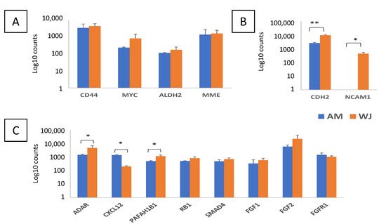
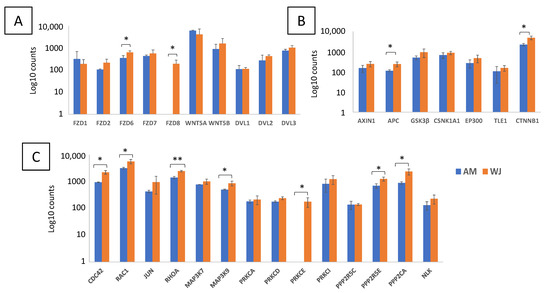
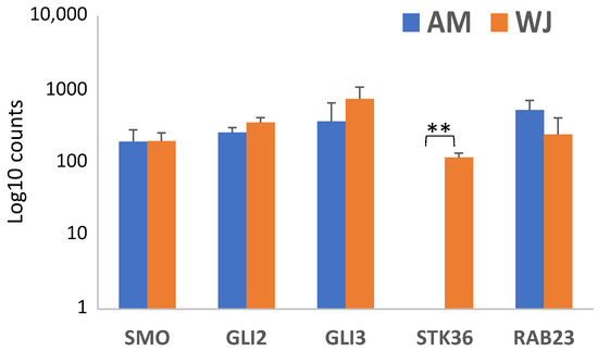
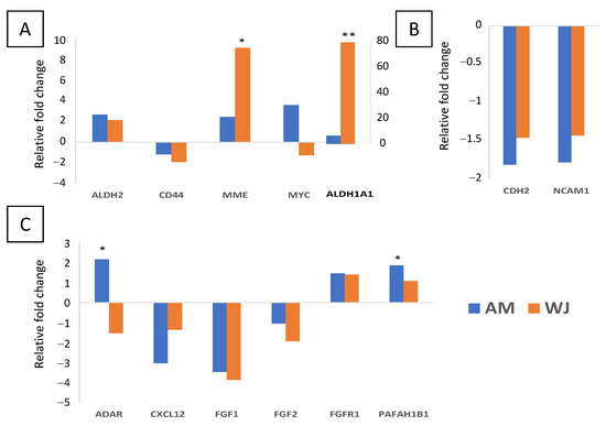
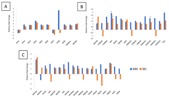
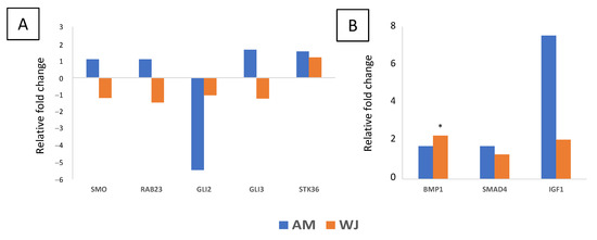
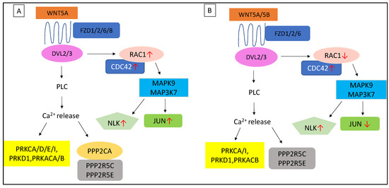
The use of perinatal mesenchymal stem cells (MSCs) in bone tissue regeneration and engineering to substitute bone marrow MSCs has drawn great interest due to their high yield, ease of procurement, multilineage differentiation potential and lack of ethical concerns. Although amniotic membrane (AM) and Wharton’s jelly (WJ)-derived MSCs have been widely shown to possess osteogenic differentiation potential, the intrinsic properties determining their osteogenic capacity remain unclear. Here, we compared gene expression profiles of AM- and WJ-MSCs at basal and osteogenic conditions by using the NanoString Stem Cell Panel containing regulatory genes associated with stemness, self-renewal, Wnt, Notch and Hedgehog signalling pathways. At basal condition, WJ-MSCs displayed higher expression in most genes regardless of their functional roles in self-renewal, adhesion, or differentiation signalling pathways. After osteo-induction, elevated expression of self-renewal genes ADAR and PAFAH1B1 was observed in AM-MSCs, while stemness genes MME and ALDH1A1 were upregulated in WJ-MSC. Both MSCs showed differences in genes associated with ligands, receptors and ubiquitin ligases of the Notch pathway. In addition, further evidence was demonstrated in some signalling molecules including CTBPs, protein kinases, phosphatases, RHOA, RAC1. Downstream targets HES1 and JUN especially showed higher expression in non-induced WJ-MSCs. Hedgehog genes initially expressed in both MSCs were downregulated in WJ-MSCs during osteogenesis. This study has provided insights into the intrinsic biological differences that may lead to their discrimination in therapeutic intervention.
Full article
by
Priya Subramani, Jaianand Kannaiyan, Saurabh Khare, Paulraj Balaji, Atif Abdulwahab A. Oyouni, Saad Ali S. Aljohani, Mishal Olayan Alsulami, Osama M. Al-Amer, Othman R. Alzahrani, Malik A. Altayar, Afrah Awadh Allah Alsulami and Veeramanikandan Veeramani
Ex vivo expanded decidua-basalis(DB)-derived mesenchymal stem cells (MSCs) obtained from single donors have demonstrated therapeutic benefits in in vitro and in vivo studies. In this report, the intravenous and subcutaneous administration of DB-MSCs obtained from five healthy donors was assessed considering clinical grade
[...] Read more.
Ex vivo expanded decidua-basalis(DB)-derived mesenchymal stem cells (MSCs) obtained from single donors have demonstrated therapeutic benefits in in vitro and in vivo studies. In this report, the intravenous and subcutaneous administration of DB-MSCs obtained from five healthy donors was assessed considering clinical grade proliferation, accessibility, and toxic effects in Wistar albino rats. The ability of the obtained DB-MSCs for differentiating, as well as their expression of several cell surface markers and immunomodulatory activities, were all assessed. Clinical standard proliferated cells were administered to animals intravenously and subcutaneously in a series of preclinical models in order to assess their in vivo toxicity, general safety, and tumorigenic possibilities. We established that DB cells exhibit structural and functional traits with MSCs. At various doses supplied intravenously or subcutaneously, the research showed no fatality, abnormal response to therapy, or substantial pathological modifications in the rats. Furthermore, there was no indication of prenatal damage in the same animal species when the rats were repeatedly treated with DBMSCs. Thus, DBMSCs were demonstrated to be non-toxic, non-teratogenic, and non-tumorigenic. To determine whether they can be administrated to human patients without risk, more investigation is recommended.
Full article
by
Irina V. Kholodenko, Alisa M. Gisina, Garik V. Manukyan, Alexander G. Majouga, Elena V. Svirshchevskaya, Roman V. Kholodenko and Konstantin N. Yarygin
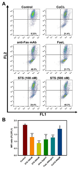
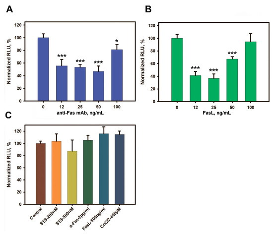
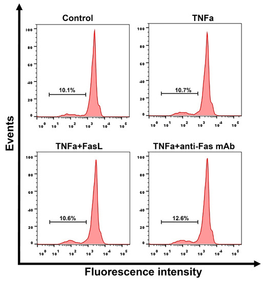
Mesenchymal stem cells (MSCs) have a pronounced therapeutic potential in various pathological conditions. Though therapeutic effects of MSC transplantation have been studied for a long time, the underlying mechanisms are still not clear. It has been shown that transplanted MSCs are rapidly eliminated,
[...] Read more.
Mesenchymal stem cells (MSCs) have a pronounced therapeutic potential in various pathological conditions. Though therapeutic effects of MSC transplantation have been studied for a long time, the underlying mechanisms are still not clear. It has been shown that transplanted MSCs are rapidly eliminated, presumably by apoptosis. As the mechanisms of MSC apoptosis are not fully understood, in the present work we analyzed MSC sensitivity to Fas-induced apoptosis using MSCs isolated from the biopsies of liver fibrosis patients (L-MSCs). The level of cell death was analyzed by flow cytometry in the propidium iodide test. The luminescent ATP assay was used to measure cellular ATP levels; and the mitochondrial membrane potential was assessed using the potential-dependent dye JC-1. We found that human L-MSCs were resistant to Fas-induced cell death over a wide range of FasL and anti-Fas mAb concentrations. At the same time, intrinsic death signal inducers CoCl2 and staurosporine caused apoptosis of L-MSCs in a dose-dependent manner. Despite the absence of Fas-induced cell death treatment of L-MSCs with low concentrations of FasL or anti-Fas mAb resulted in a cellular ATP level decrease, while high concentrations of the inducers caused a decline of the mitochondrial membrane potential. Pre-incubation of L-MSCs with the pro-inflammatory cytokine TNF-α did not promote L-MSC cell death. Our data indicate that human L-MSCs have increased resistance to receptor-mediated cell death even under inflammatory conditions.
Full article
Spinal cord injury (SCI) is a destructive condition that results in lasting neurological damage resulting in disruption of the connection between the central nervous system and the rest of the body. Currently, there are several approaches in the treatment of a damaged spinal
[...] Read more.
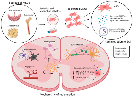
Spinal cord injury (SCI) is a destructive condition that results in lasting neurological damage resulting in disruption of the connection between the central nervous system and the rest of the body. Currently, there are several approaches in the treatment of a damaged spinal cord; however, none of the methods allow the patient to return to the original full-featured state of life before the injury. Cell transplantation therapies show great potential in the treatment of damaged spinal cords. The most examined type of cells used in SCI research are mesenchymal stromal cells (MSCs). These cells are at the center of interest of scientists because of their unique properties. MSCs regenerate the injured tissue in two ways: (i) they are able to differentiate into some types of cells and so can replace the cells of injured tissue and (ii) they regenerate tissue through their powerful known paracrine effect. This review presents information about SCI and the treatments usually used, aiming at cell therapy using MSCs and their products, among which active biomolecules and extracellular vesicles predominate.
Full article
Over the past two decades, mesenchymal stem cells (MSCs) have shown promising therapeutic effects both in preclinical studies (in animal models of a wide range of diseases) and in clinical trials. However, the efficacy of MSC-based therapy is not always predictable. Moreover, despite
[...] Read more.
Over the past two decades, mesenchymal stem cells (MSCs) have shown promising therapeutic effects both in preclinical studies (in animal models of a wide range of diseases) and in clinical trials. However, the efficacy of MSC-based therapy is not always predictable. Moreover, despite the large number of studies, the mechanisms underlying the regenerative potential of MSCs are not fully elucidated. Recently, it has been reliably established that transplanted MSCs can undergo rapid apoptosis and clearance from the recipient’s body, still exhibiting therapeutic effects, especially those associated with their immunosuppressive/immunomodulating properties. The mechanisms underlying these effects can be mediated by the efferocytosis of apoptotic MSCs by host phagocytic cells. In this concise review, we briefly describe three types of MSC-generated extracellular vesicles, through which their therapeutic functions can potentially be carried out; we focused on reviewing recent data on apoptotic MSCs and MSC-derived apoptotic bodies (MSC-ApoBDs), their functions, and the mechanisms of their therapeutic effects.
Full article
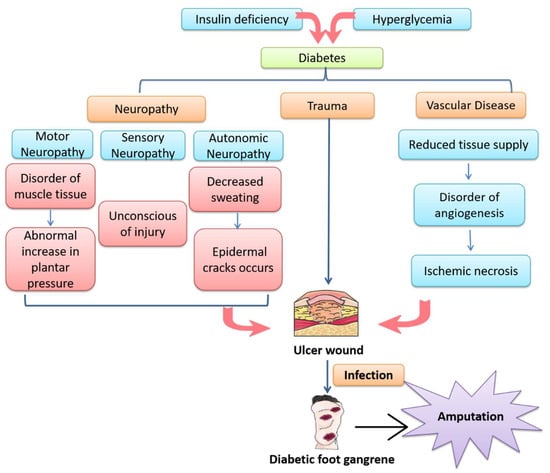
Impaired healing of diabetic wounds harms patients’ quality of life and even leads to disability and death, which is an urgent issue to be solved clinically. Despite the great progress that has been achieved, it remains a worldwide challenge to develop effective therapeutic
[...] Read more.
Impaired healing of diabetic wounds harms patients’ quality of life and even leads to disability and death, which is an urgent issue to be solved clinically. Despite the great progress that has been achieved, it remains a worldwide challenge to develop effective therapeutic treatments for diabetic wounds. Recently, exosomes have attracted special attention because they can be involved in immune response, antigen presentation, cell migration, cell differentiation, tumor invasion and other processes. Meanwhile, exosomes have been proven to hold great potential in the treatment of diabetic wounds. Mechanistic studies of exosomes based on signaling pathways could not only help to uncover the mechanisms by which exosomes promote diabetic wound healing but could also provide a theoretical basis for the clinical application of exosomes. Herein, our mini-review aims to summarize the progress of research on the use of various exosomes derived from different cell types to promote diabetic wound healing, with a focus on the classical signaling pathways, including PI3K/Akt, Wnt, NF-κB, MAPK, Notch, Nrf2, HIF-1α/VEGF and TGF-β/Smad. The results show that exosomes could regulate these signaling pathways to down-regulate inflammation, reduce oxidative stress, increase angiogenesis, promote fibroblast proliferation, induce re-epithelization and inhibit scar formation, making exosomes attractive candidates for the treatment of diabetic wounds.
Full article


