Sign in to use this feature.

Years

Between: -

Article Types

Countries / Regions

Search Results (41)

Search Parameters:
Journal = Organoids

Order results
Result details
Results per page
Select all
Export citation of selected articles as:
22 pages, 4970 KiB  
Article
Single-Cell Assessment of Human Stem Cell-Derived Mesolimbic Models and Their Responses to Substances of Abuse
by Thomas P. Rudibaugh, Ryan W. Tam, R. Chris Estridge, Samantha R. Stuppy and Albert J. Keung
Organoids 2024, 3(2), 126-147; https://doi.org/10.3390/organoids3020009 - 20 Jun 2024
Viewed by 288
Abstract
The mesolimbic pathway connects ventral tegmental area dopaminergic neurons and striatal medium spiny neurons, playing a critical role in reward and stress behaviors. Exposure to substances of abuse during development and adulthood has been linked to adverse outcomes and molecular changes. The rise [...] Read more.
The mesolimbic pathway connects ventral tegmental area dopaminergic neurons and striatal medium spiny neurons, playing a critical role in reward and stress behaviors. Exposure to substances of abuse during development and adulthood has been linked to adverse outcomes and molecular changes. The rise of human cell repositories and whole-genome sequences enables human functional genomics ‘in a dish’, offering insights into human-specific responses to substances of abuse. Continued development of new models is needed, and the characterization of in vitro models is also necessary to ensure appropriate experimental designs and the accurate interpretation of results. This study introduces new culture conditions for generating medium spiny neurons and dopaminergic neurons with an early common media, allowing for coculture and assembloid generation. It then provides a comprehensive characterization of these and prior models and their responses to substances of abuse. Single-cell analysis reveals cell-type-specific transcriptomic responses to dopamine, cocaine, and morphine, including compound and cell-type-specific transcriptomic signatures related to neuroinflammation and alterations in signaling pathways. These findings offer a resource for future genomics studies leveraging human stem cell-derived models. Full article
(This article belongs to the Special Issue The Current Applications and Potential of Stem Cell-Derived Organoids)
Show Figures
Figure 1

---

Figure 3

---

Figure 4

---

Figure 5

13 pages, 2038 KiB  
Protocol
Development and Optimization of a Lactate Dehydrogenase Assay Adapted to 3D Cell Cultures
by Héloïse Castiglione, Lucie Madrange, Thomas Lemonnier, Jean-Philippe Deslys, Frank Yates and Pierre-Antoine Vigneron
Organoids 2024, 3(2), 113-125; https://doi.org/10.3390/organoids3020008 - 5 Jun 2024
Viewed by 584
Abstract
In recent years, 3D cell culture systems have emerged as sophisticated in vitro models, providing valuable insights into human physiology and diseases. The transition from traditional 2D to advanced 3D cultures has introduced novel obstacles, complicating the characterization and analysis of these models. [...] Read more.
In recent years, 3D cell culture systems have emerged as sophisticated in vitro models, providing valuable insights into human physiology and diseases. The transition from traditional 2D to advanced 3D cultures has introduced novel obstacles, complicating the characterization and analysis of these models. While the lactate dehydrogenase (LDH) activity assay has long been a standard readout for viability and cytotoxicity assessments in 2D cultures, its applicability in long-term 3D cultures is hindered by inappropriate normalization and low LDH stability over time. In response to these challenges, we propose an optimization of LDH assays, including a crucial normalization step based on total protein quantification and a storage method using an LDH preservation buffer. We applied it to compare unexposed cerebral organoids with organoids exposed to a toxic dose of valproic acid, and showed efficient normalization of cellular viability as well as enhanced LDH stability within the buffer. Importantly, normalized LDH activity results obtained were independent of organoid dimension and cell density. This refined LDH assay, tailored to address 3D culture constraints, allows for the transposition of this routine test from 2D to 3D cultures. Full article
Show Figures
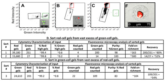
Figure 1

---

Figure 3

---

Figure 4

30 pages, 1802 KiB  
Communication
Organoids, Biocybersecurity, and Cyberbiosecurity—A Light Exploration
by Xavier Palmer, Cyril Akafia, Eleasa Woodson, Amanda Woodson and Lucas Potter
Organoids 2024, 3(2), 83-112; https://doi.org/10.3390/organoids3020007 - 13 May 2024
Viewed by 1051
Abstract
Organoids present immense promise for studying organ systems and their functionality. Recently, they have become the subject of exploration outside of purely biomedical uses in multiple directions. We will explore the rapidly evolving landscape of organoid research over the 21st century, discussing significant [...] Read more.
Organoids present immense promise for studying organ systems and their functionality. Recently, they have become the subject of exploration outside of purely biomedical uses in multiple directions. We will explore the rapidly evolving landscape of organoid research over the 21st century, discussing significant advancements in organoid research and highlighting breakthroughs, methodologies, and their transformative impact on our understanding of physiology and modeling. In addition, we will explore their potential use for biocomputing and harnessing organoid intelligence, investigate how these miniaturized organ-like structures promise to create novel computational models and processing platforms allowing for innovative approaches in drug discovery, personalized medicine, and disease prediction. Lastly, we will address the ethical dilemmas surrounding organoid research by dissecting the intricate ethical considerations related to the creation, use, and potential implications of these in vitro models. Through this work, the goal of this paper is to provide introductory perspectives and bridges that will connect organoids to cybersecurity applications and the imperative ethical discourse accompanying its advancements with commentary on future uses. Full article
Show Figures
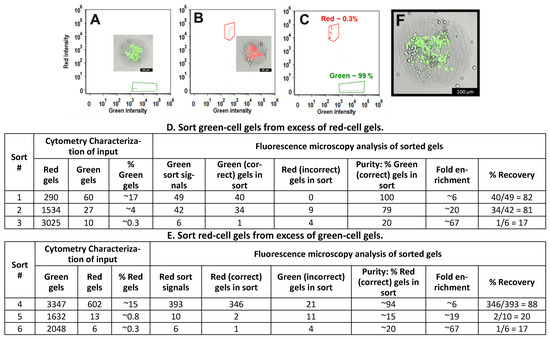
Figure 1

---

Figure 3

---

Figure 4

---

Figure 5

16 pages, 1673 KiB  
Review
Treatment of Canine Type 1 Diabetes Mellitus: The Long Road from Twice Daily Insulin Injection towards Long-Lasting Cell-Based Therapy
by Flavia C. M. Oliveira, Annemarie W. Y. Voorbij, Elisa C. Pereira, Leonor M. M. Alves e Almeida, Geanne R. Moraes, Joana T. De Oliveira, Boyd H. T. Gouw, Sabrina A. M. Legatti, Hans S. Kooistra, Bart Spee, Andre M. C. Meneses and Louis C. Penning
Organoids 2024, 3(2), 67-82; https://doi.org/10.3390/organoids3020006 - 4 Apr 2024
Viewed by 1107
Abstract
For over 150 years, researchers have studied the (patho)physiology of the endocrine pancreas and devised treatment options for diabetes mellitus (DM). However, no cure has been developed so far. In dogs, diabetes mellitus type 1 (T1DM) is the most common presentation. Treatment consists [...] Read more.
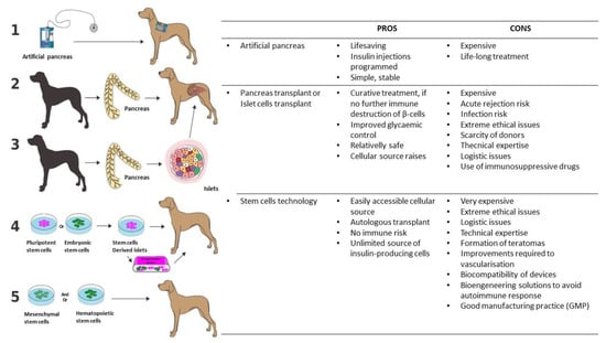
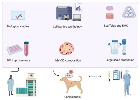
For over 150 years, researchers have studied the (patho)physiology of the endocrine pancreas and devised treatment options for diabetes mellitus (DM). However, no cure has been developed so far. In dogs, diabetes mellitus type 1 (T1DM) is the most common presentation. Treatment consists of twice daily insulin injections, monitored by spatial blood glucose measurements. Even though dogs were instrumental in the discovery of insulin and islet transplantations, the treatment in diabetic dogs has remained unchanged for decades. Providing twice daily insulin injections is demanding for both owners and dogs and may result in hypoglycaemic events, creating the need for new treatment strategies. Novel regenerative medicine-based tools, such as improved β-cell culture protocols and artificial devices, have sparked hope for a cure. In human medicine, emerging technologies such as the transplantation of insulin-producing β-cells, generated by stem cell differentiation, with or without an encapsulation device, are currently tested in phase I/II clinical trials. As the pathogenesis of T1DM is remarkably similar between humans and dogs, novel treatment methods could be implemented in canine medicine. This review briefly summarises the physiology of the canine endocrine pancreas and the pathophysiology of canine DM before exploring current and possible future treatment options for canine DM. Full article
Show Figures
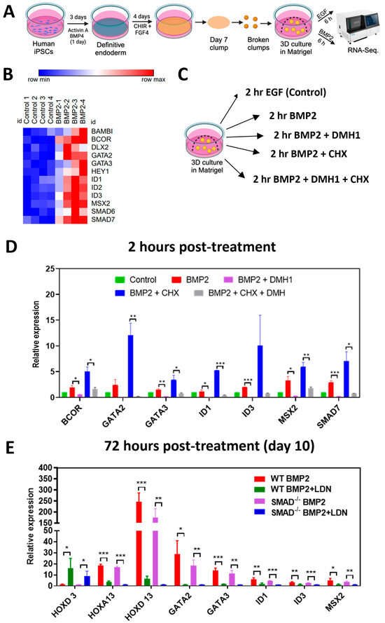
Figure 1

---

Figure 3

---

Figure 4

13 pages, 3731 KiB  
Protocol
Generation of Trophoblast Organoids from Chorionic Villus Sampling
by Bas van Rijn, Diane Van Opstal, Nicole van Koetsveld, Maarten Knapen, Joost Gribnau and Olivier Schäffers
Organoids 2024, 3(1), 54-66; https://doi.org/10.3390/organoids3010005 - 5 Mar 2024
Cited by 1 | Viewed by 1083
Abstract
Studying human placental development and function presents significant challenges due to the inherent difficulties in obtaining and maintaining placental tissue throughout the course of an ongoing pregnancy. Here, we provide a detailed protocol for generating trophoblast organoids from chorionic villi obtained during ongoing [...] Read more.
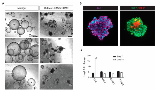
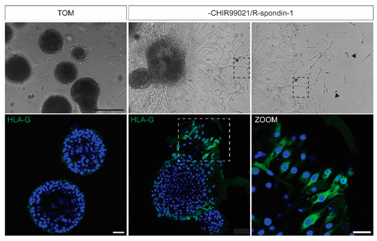
Studying human placental development and function presents significant challenges due to the inherent difficulties in obtaining and maintaining placental tissue throughout the course of an ongoing pregnancy. Here, we provide a detailed protocol for generating trophoblast organoids from chorionic villi obtained during ongoing pregnancy. Our method results in efficient generation of trophoblast organoids from chorionic villus sampling, does not require preselection of chorionic villi, and controls contamination of decidual gland organoids. The resulting trophoblast organoids spontaneously form syncytiotrophoblasts that start secreting hCG hormone amongst other placenta-specific factors. Our approach facilitates the generation of trophoblast organoids from a variety of genetic backgrounds, including trisomies and gene mutations, and can be aligned with prenatal diagnostic routines. The protocol requires up to 14 days and can be carried out by users with expertise in cell culture. Full article
Show Figures
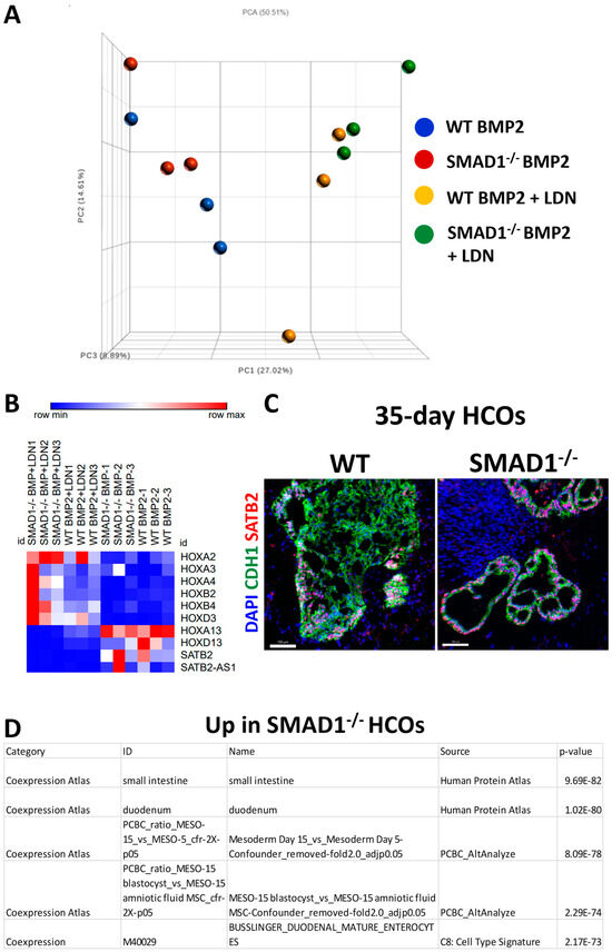
Figure 1

---

Figure 3

---

Figure 4

---

Figure 5

19 pages, 50653 KiB  
Article
Analysis of Osteosarcoma Cell Lines and Patient Tissue Using a 3D In Vivo Tumor Model—Possible Effects of Punicalagin
by Anna Rebecca Dorn, Sara Neff, Sophia Hupp, Melissa Engelhardt, Eric Pion, Ulrich Lenze, Carolin Knebel, Anna Duprée, Simone Schewe, Markus Weber, Christian Wulbrand, Axel Hillmann, Florian Weber, Phillip Clarke, Philipp Kainz, Thiha Aung and Silke Haerteis
Organoids 2024, 3(1), 35-53; https://doi.org/10.3390/organoids3010004 - 4 Mar 2024
Viewed by 1393
Abstract
Osteosarcomas are the most common primary malignant bone tumors and mostly affect children, adolescents, and young adults. Despite current treatment options such as surgery and polychemotherapy, the survival of patients with metastatic disease remains poor. In recent studies, punicalagin has reduced the cell [...] Read more.
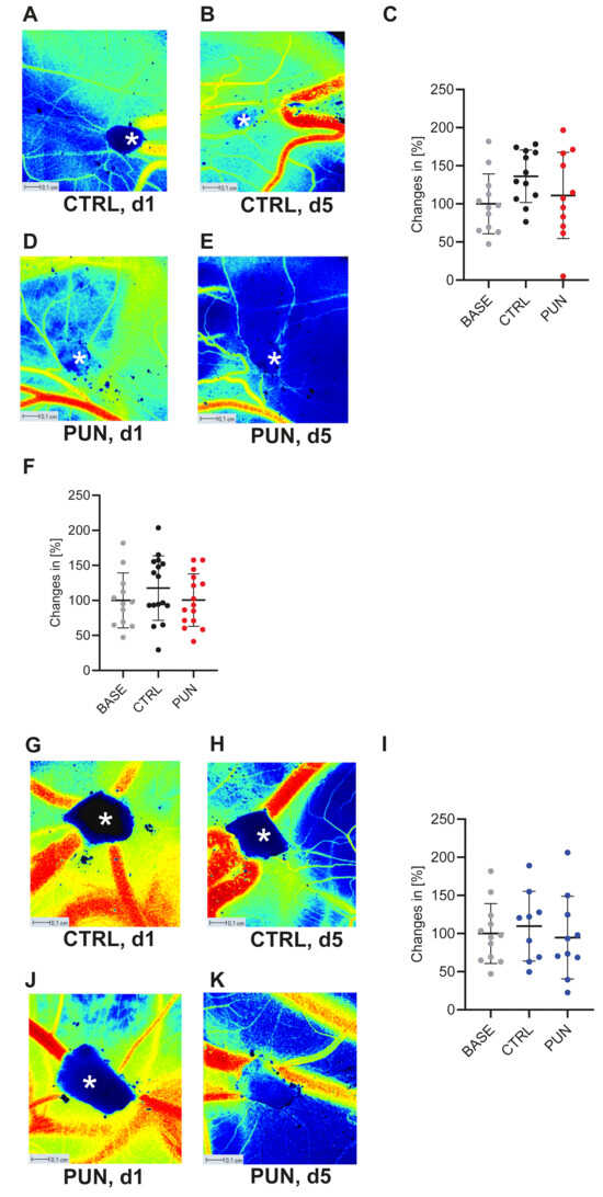
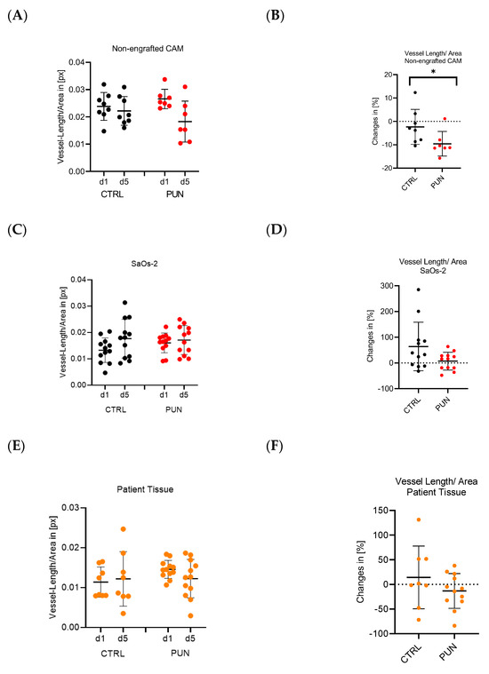
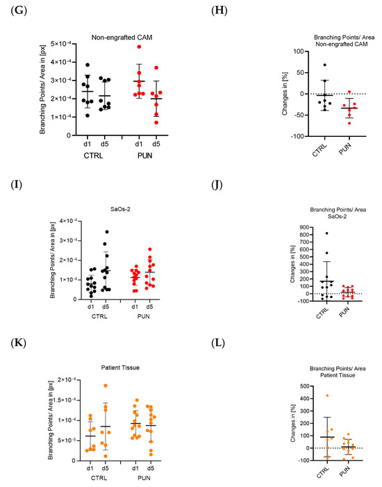
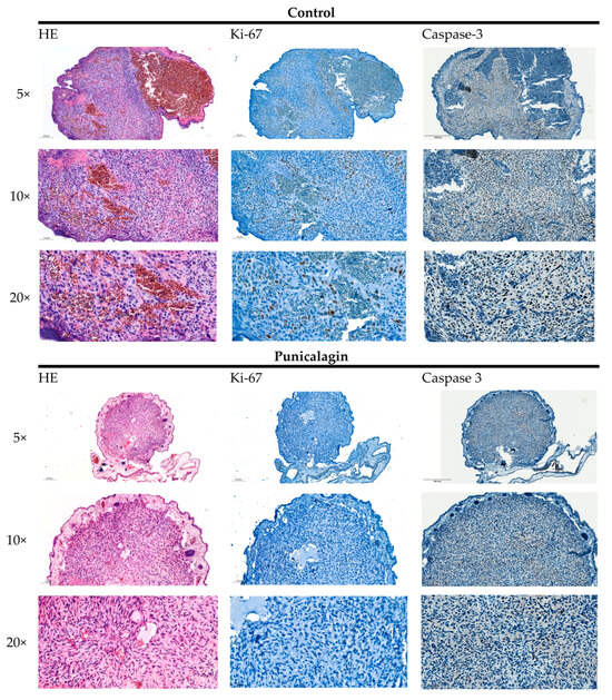
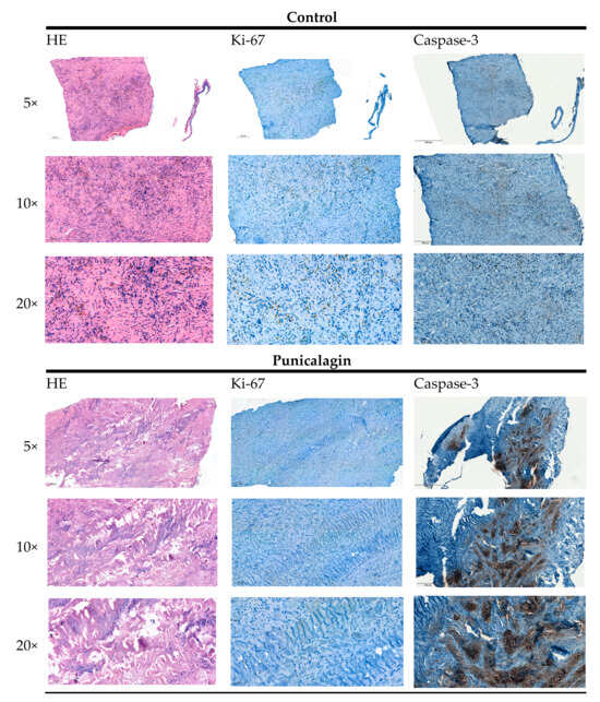
Osteosarcomas are the most common primary malignant bone tumors and mostly affect children, adolescents, and young adults. Despite current treatment options such as surgery and polychemotherapy, the survival of patients with metastatic disease remains poor. In recent studies, punicalagin has reduced the cell viability, angiogenesis, and invasion in cell culture trials. The aim of this study was to examine the effects of punicalagin on osteosarcomas in a 3D in vivo tumor model. Human osteosarcoma biopsies and SaOs-2 and MG-63 cells, were grown in a 3D in vivo chorioallantoic membrane (CAM) model. After a cultivation period of up to 72 h, the tumors received daily treatment with punicalagin for 4 days. Weight measurements of the CAM tumors were performed, and laser speckle contrast imaging (LSCI) and a deep learning-based image analysis software (CAM Assay Application v.3.1.0) were used to measure angiogenesis. HE, Ki-67, and Caspase-3 staining was performed after explantation. The osteosarcoma cell lines SaOs-2 and MG-63 and osteosarcoma patient tissue displayed satisfactory growth patterns on the CAM. Treatment with punicalagin decreased tumor weight, proliferation, and tumor-induced angiogenesis, and the tumor tissue showed pro-apoptotic characteristics. These results provide a robust foundation for the implementation of further studies and show that punicalagin offers a promising supplementary treatment option for osteosarcoma patients. The 3D in vivo tumor model represents a beneficial model for the testing of anti-cancer therapies. Full article
Show Figures
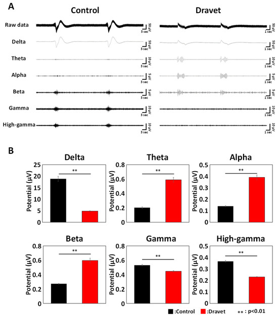
Figure 1

---

Figure 3

---

Figure 4

---

Figure 4 Cont.

---

Figure 5

---

Figure 6

3 pages, 159 KiB  
Editorial
The Next Generation of Organoids Will Be More Complex and Even Closer to Resembling Real Organs: An Interview with Prof. Dr. Hans Clevers
by Süleyman Ergün and Organoids Editorial Office
Organoids 2024, 3(1), 32-34; https://doi.org/10.3390/organoids3010003 - 20 Feb 2024
Viewed by 876
Abstract
In this issue, we are pleased and honored to have an interview with Professor Hans Clevers, who is the Advisory Board Member of Organoids [...] Full article
14 pages, 2347 KiB  
Article
Human Nasal Epithelium Organoids for Assessing Neutralizing Antibodies to a Protective SARS-CoV-2 Virus-like Particle Vaccine
by Julio Carrera Montoya, Simon Collett, Daniel Fernandez Ruiz, Linda Earnest, Melissa A. Edeling, Ashley Huey Yiing Yap, Chinn Yi Wong, James P. Cooney, Kathryn C. Davidson, Jason Roberts, Steven Rockman, Bang M. Tran, Julie L. McAuley, Georgia Deliyannis, Samantha L. Grimley, Damian F. J. Purcell, Shafagh A. Waters, Dale I. Godfrey, Dhiraj Hans, Marc Pellegrini, Jason M. Mackenzie, Elizabeth Vincan, William R. Heath and Joseph Torresiadd Show full author list remove Hide full author list
Organoids 2024, 3(1), 18-31; https://doi.org/10.3390/organoids3010002 - 1 Feb 2024
Viewed by 1501
Abstract
Existing mRNA COVID-19 vaccines have shown efficacy in reducing severe cases and fatalities. However, their effectiveness against infection caused by emerging SARS-CoV-2 variants has waned considerably, necessitating the development of variant vaccines. Ideally, next-generation vaccines will be capable of eliciting broader and more [...] Read more.
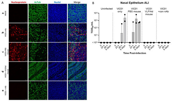
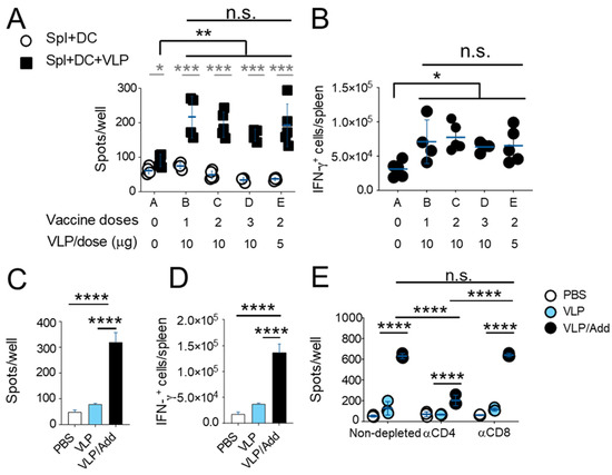
Existing mRNA COVID-19 vaccines have shown efficacy in reducing severe cases and fatalities. However, their effectiveness against infection caused by emerging SARS-CoV-2 variants has waned considerably, necessitating the development of variant vaccines. Ideally, next-generation vaccines will be capable of eliciting broader and more sustained immune responses to effectively counteract new variants. Additionally, in vitro assays that more closely represent virus neutralization in humans would greatly assist in the analysis of protective vaccine-induced antibody responses. Here, we present findings from a SARS-CoV-2 VLP vaccine encompassing three key structural proteins: Spike (S), Envelope (E), and Membrane (M). The VLP vaccine effectively produced neutralizing antibodies as determined by surrogate virus neutralization test, and induced virus-specific T-cell responses: predominantly CD4+, although CD8+ T cell responses were detected. T cell responses were more prominent with vaccine delivered with AddaVax compared to vaccine alone. The adjuvanted vaccine was completely protective against live virus challenge in mice. Furthermore, we utilized air–liquid-interface (ALI)-differentiated human nasal epithelium (HNE) as an in vitro system, which authentically models human SARS-CoV-2 infection and neutralization. We show that immune sera from VLP-vaccinated mice completely neutralized SARS-CoV-2 virus infection, demonstrating the potential of ALI-HNE to assess vaccine induced Nab. Full article
Show Figures
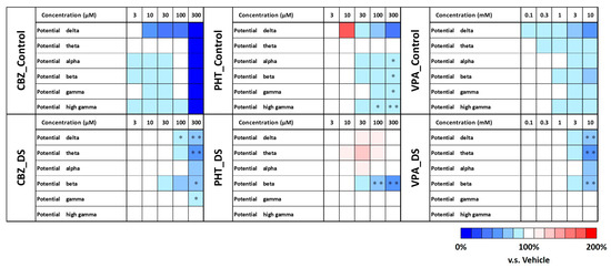
Figure 1

---

Figure 3

---

Figure 4

17 pages, 12196 KiB  
Article
Visualization of Vascular Perfusion of Human Pancreatic Cancer Tissue in the CAM Model and Its Impact on Future Personalized Drug Testing
by Andreas Ettner-Sitter, Agata Montagner, Jonas Kuenzel, Kathrin Brackmann, Maximilian Schäfer, Robert Schober, Florian Weber, Thiha Aung, Christina Hackl and Silke Haerteis
Organoids 2024, 3(1), 1-17; https://doi.org/10.3390/organoids3010001 - 8 Jan 2024
Cited by 1 | Viewed by 1460
Abstract
Although significant improvements have been made in the treatment of pancreatic cancer, its prognosis remains poor with an overall 5-year survival rate of less than 10%. New experimental approaches are necessary to develop novel therapeutics. In this study, the investigation of pancreatic cancer [...] Read more.
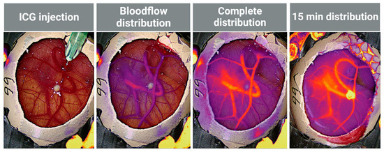
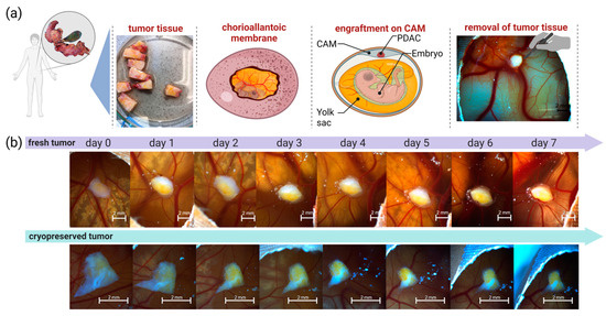
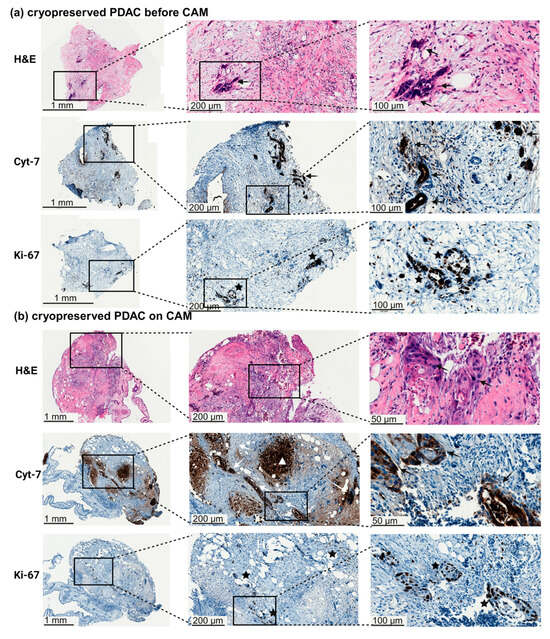
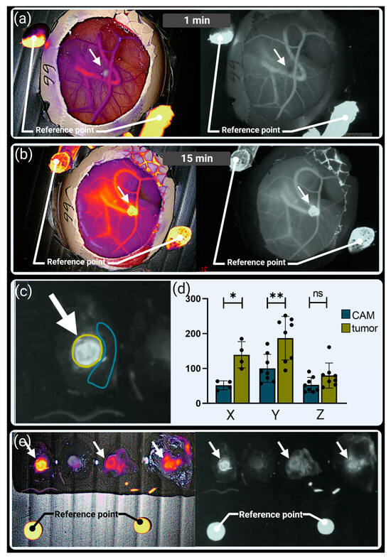
Although significant improvements have been made in the treatment of pancreatic cancer, its prognosis remains poor with an overall 5-year survival rate of less than 10%. New experimental approaches are necessary to develop novel therapeutics. In this study, the investigation of pancreatic cancer tissue growth in the chorioallantoic membrane (CAM) model and the subsequent use of indocyanine green (ICG) injections for the verification of intratumoral perfusion was conducted. ICG was injected into the CAM vasculature to visualize the perfusion of the tumor tissue. The presence of metastasis was investigated through PCR for the human-specific ALU element in the liver of the chicken embryo. Additionally, the usage of cryopreserved pancreatic tumors was established. Intratumoral perfusion of tumor tissue on the CAM was observed in recently obtained and cryopreserved tumors. ALU-PCR detected metastasis in the chick embryos’ livers. After cryopreservation, the tissue was still vital, and the xenografts generated from these tumors resembled the histological features of the primary tumor. This methodology represents the proof of principle for intravenous drug testing of pancreatic cancer in the CAM model. The cryopreserved tumors can be used for testing novel therapeutics and can be integrated into the molecular tumor board, facilitating personalized tumor treatment. Full article
Show Figures

Graphical abstract

---

Figure 2

---

Figure 3

---

Figure 4

---

Figure 5

---

Figure 6

---

Figure 7

---

Figure 8

17 pages, 2418 KiB  
Review
Vascularizing Organoids to Promote Long-Term Organogenesis on a Chip
by Xinhui Wang, Brent M. Bijonowski and Nicholas A. Kurniawan
Organoids 2023, 2(4), 239-255; https://doi.org/10.3390/organoids2040019 - 7 Dec 2023
Viewed by 2296
Abstract
Organoids have emerged as a powerful tool for studying organ development, disease modeling, and drug discovery due to their ability to mimic the in vivo structure and function of organs in a three-dimensional in vitro model. During in vivo organ maturation, the process [...] Read more.
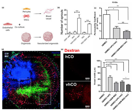
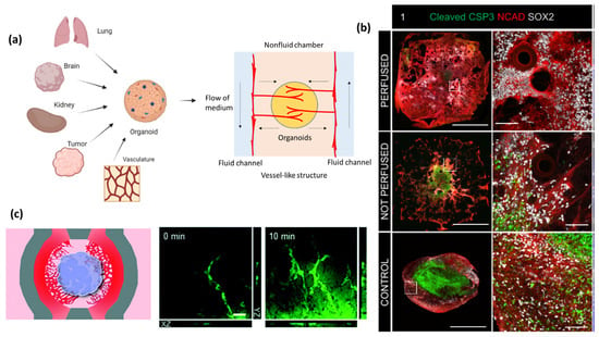
Organoids have emerged as a powerful tool for studying organ development, disease modeling, and drug discovery due to their ability to mimic the in vivo structure and function of organs in a three-dimensional in vitro model. During in vivo organ maturation, the process of vascularization is crucial for the provision of nutrients and oxygen to cells and the removal of waste products as the organ increases in size. Similarly, organoids can grow to sizes greater than the millimeter scale, yet transport of oxygen and nutrients to the center becomes increasingly difficult, often resulting in the formation of a necrotic core. Herein, we provide a concise summary of the recent development of methods to initiate and maintain vascularization of organoids. Broadly, vascularization of organoids has been achieved primarily by two means: generating organoids that contain endothelial cells or employing the secretion of vascular growth factors to promote vascularization. Growth factors play a fundamental role in regulating blood vessel formation through chemical signals that cause changes in the cell–cell adhesions and ultimately the migration of endothelial cells. Furthermore, models with perfusable systems demonstrate that through the application of growth factors and cells, the vascular network in vascularization-based organoids can administer biological substances to the interior of the organoid, opening up new possibilities for long-term organoid culture in vitro. This goal is being realized through the development of bioengineering tools, such as vascularized organoids on a chip, which are currently tested for various organ systems, including the lung, brain, kidney, and tumors, with applications in cancer angiogenesis and metastasis research. Taken together, our review underlines the vast potential of vascularized organoids to improve the understanding of organ development, while also proposing exciting avenues of organoid-on-a-chip and disease modeling. Full article
Show Figures

Graphical abstract

---

Figure 2

---

Figure 3

---

Figure 4

8 pages, 247 KiB  
Commentary
Stem Cell-Derived Organoids, Embryoids, and Embryos: Advances in Organismic Development In Vitro Force Us to Re-Focus on Ethical and Legal Aspects of Model Choice
by Hans-Werner Denker
Organoids 2023, 2(4), 231-238; https://doi.org/10.3390/organoids2040018 - 5 Dec 2023
Viewed by 1026
Abstract
While research on stem cell-derived tissues and organoids is rapidly expanding, the technically related creation of complex embryoids has recently excited a vivid discussion since it raises ethical questions about individuation and the possible gain of viability. The present study focuses on the [...] Read more.
While research on stem cell-derived tissues and organoids is rapidly expanding, the technically related creation of complex embryoids has recently excited a vivid discussion since it raises ethical questions about individuation and the possible gain of viability. The present study focuses on the onset of organismic development and the proposed biological and legal definitions for the terms embryo, embryoid, and organoid. It is concluded that such considerations have become important for investigators’ choices of the appropriate in vitro model systems, allowing the formation of organoids vs. complex embryoids. Full article
13 pages, 352 KiB  
Review
Modelling Meningioma Using Organoids: A Review of Methodologies and Applications
by Clara Elena López Vásquez, Clint Gray, Claire Henry and Matthew J. Munro
Organoids 2023, 2(4), 218-230; https://doi.org/10.3390/organoids2040017 - 4 Dec 2023
Viewed by 1524
Abstract
Meningiomas are the most common tumours of the central nervous system. According to the World Health Organization (WHO), this disease is classified into three different grades: 80% of meningioma patients present with benign grade I tumours, while less than 2% present with malignant [...] Read more.
Meningiomas are the most common tumours of the central nervous system. According to the World Health Organization (WHO), this disease is classified into three different grades: 80% of meningioma patients present with benign grade I tumours, while less than 2% present with malignant grade III meningiomas. Despite affecting thousands of people worldwide, much remains unknown about this disease, and the development of systemic treatments is still far behind in comparison to other types of tumours. Therefore, forming 3D structures (spheroids and organoids) could facilitate research on the mechanisms of formation, proliferation, migration, and invasion of these, for the most part, benign tumours, while also helping in the process of drug development. To date, there are three published methods for the formation of meningioma organoids primarily derived from patient tissue samples. Organoids offer many advantages in the development of treatments because they recapitulate the cellular complexity within tumours. These new methodological advances could open a substantial number of possibilities for the further characterisation and treatment of meningiomas. This review includes an overview of the disease and a description and comparison of established protocols for meningioma organoid formation. Full article
14 pages, 3802 KiB  
Article
The Rapid Generation of Cell-Laden, FACS-Compatible Collagen Gels
by Yi Xiao, Qiaoling Huang, Jesse W. Collins, Julie Brouchon, Jeffery A. Nelson, Zachary Niziolek, Alison O’Neil, Fangfu Ye, David A. Weitz and John A. Heyman
Organoids 2023, 2(4), 204-217; https://doi.org/10.3390/organoids2040016 - 17 Nov 2023
Cited by 1 | Viewed by 1337
Abstract
A three-dimensional cell culture in hydrogel beads can support cell growth and differentiation into multi-cellular structures, and these gel beads could be used as building blocks for more complex three-dimensional assemblies. This requires hydrogel beads that are robust enough to sort via FACS [...] Read more.
A three-dimensional cell culture in hydrogel beads can support cell growth and differentiation into multi-cellular structures, and these gel beads could be used as building blocks for more complex three-dimensional assemblies. This requires hydrogel beads that are robust enough to sort via FACS yet can be degraded by cell-secreted enzymes. Collagen polymers form hydrogels that are excellent cell growth substrates; however, collagen-containing hydrogel beads typically include additional polymers that limit their degradation. Here, we introduce a simple microfluidic method to generate robust, sortable, cell-laden collagen hydrogel beads. We use on-device pH control to trigger collagen gelation without exposing cells to low pH, ensuring high cell viability. We fabricate microfluidic devices to generate droplets with a wide size range, as demonstrated by production of both small (~55 µm diameter) and large (~300 µm diameter) collagen gels. All hydrogels are sufficiently robust to allow for sorting using FACS. Moreover, high cell viability is maintained throughout the process. Full article
Show Figures

Graphical abstract

---

Figure 2

---

Figure 3

---

Figure 4

---

Figure 5

---

Figure 6

12 pages, 4377 KiB  
Article
SMAD1 Is Dispensable for CDX2 Induction but Required for the Repression of Ectopic Small-Intestinal Gene Expression in Human-Pluripotent-Stem-Cell-Derived Colonic Organoids
by Na Qu, Abdelkader Daoud, Braxton Jeffcoat and Jorge O. Múnera
Organoids 2023, 2(4), 192-203; https://doi.org/10.3390/organoids2040015 - 14 Nov 2023
Viewed by 1073
Abstract
The generation of gastrointestinal tissues from human pluripotent stem cells has provided unprecedented insight into the molecular mechanisms that drive the patterning of the primitive gut tube. Previous work has identified bone-morphogenetic-protein (BMP) signaling as an important mediator of mid/hindgut versus foregut and [...] Read more.
The generation of gastrointestinal tissues from human pluripotent stem cells has provided unprecedented insight into the molecular mechanisms that drive the patterning of the primitive gut tube. Previous work has identified bone-morphogenetic-protein (BMP) signaling as an important mediator of mid/hindgut versus foregut and hindgut versus midgut cell fate choice. Inhibition of BMP signaling during gut tube morphogenesis inhibits the expression of the pan-intestinal transcription factor CDX2. Treatment of CDX2+ mid/hindgut cultures with BMP patterns them into hindgut, which gives rise to colonic organoids (HCOs). While the role for BMP signaling is clear, the molecular mechanisms through which BMP signaling patterns the mid/hindgut and colon remain unclear. BMPs bind to BMP receptors, activating a signaling cascade that results in the activation of SMADs, which function as transcription factors. We hypothesized that one of these factors, SMAD1, would be necessary for establishing the CDX2 domain and the colon domain. Unexpectedly, endoderm derived from SMAD1-deficient induced pluripotent stem cells was capable of inducing CDX2 in response to WNT and FGF signaling. In addition, CDX2+ gut tube cultures could activate posterior HOX genes in response to BMP. However, examination of HCOs following cytodifferentiation revealed that SMAD1-deficient HCOs ectopically expressed small-intestinal markers despite expressing posterior HOX genes. These results indicate that there is redundancy of SMADs during early hindgut patterning but that SMAD1 is required for the inhibition of small-intestinal gene expression in HCOs. Full article
Show Figures

Figure 1

---

Figure 3

---

Figure 4

15 pages, 3407 KiB  
Article
Contraindicated Drug Responses in Dravet Syndrome Brain Organoids Utilizing Micro Electrode Array Assessment Methods
by Remi Yokoi, Nami Nagafuku, Yuto Ishibashi, Naoki Matsuda and Ikuro Suzuki
Organoids 2023, 2(4), 177-191; https://doi.org/10.3390/organoids2040014 - 26 Oct 2023
Viewed by 1346
Abstract
Ensuring drug safety for patients with specific neurological disorders is of paramount importance. For instance, certain antiepileptic drugs (AEDs) are contraindicated in Dravet Syndrome (DS), which is characterized by a deficiency in Na+ channel function. Constructing in vitro assessment methods capable of [...] Read more.
Ensuring drug safety for patients with specific neurological disorders is of paramount importance. For instance, certain antiepileptic drugs (AEDs) are contraindicated in Dravet Syndrome (DS), which is characterized by a deficiency in Na+ channel function. Constructing in vitro assessment methods capable of detecting contraindicated drug responses and medication effects on neurons derived from DS patients is highly anticipated for drug safety assessment and therapeutic innovation. This study used micro electrode array (MEA) measurements with low-frequency analysis on human iPSC-derived DS organoids to investigate AED responses. When exposed to the contraindicated drugs carbamazepine and phenytoin, the number of network oscillations increased in DS organoids while maintaining oscillation intensity. Furthermore, carbamazepine administration appeared to enhance activities beyond oscillations which is partially consistent with findings in the DS mouse model. Conversely, treatment with the therapeutic drug sodium valproate resulted in a similar decrease in activity both in healthy and DS organoids. The frequency characteristics of spontaneous firings and AEDs responsiveness in DS organoids demonstrated partial correlation with typical electroencephalography patterns observed in vivo. In conclusion, this study, employing MEA measurements with low-frequency analysis, revealed contraindicated drug responses and disease-specific functional characteristics in DS organoids, effective for DS patient safety assessment, precision medicine, and antiepileptic drug screening. Full article
Show Figures

Figure 1

---

Figure 3

---

Figure 4

Back to TopTop -