Int. J. Mol. Sci. 2023, 24(17), 13065; https://doi.org/10.3390/ijms241713065 - 22 Aug 2023
Cited by 6 | Viewed by 1602
Abstract
Fatty liver disease (FLD) is a clinical and pathological syndrome characterized by excessive fat deposition and even steatosis in hepatocytes. It has been proven that liver inflammation induced by fat and its derivatives are involved in the pathogenesis of FLD, while the precise
[...] Read more.
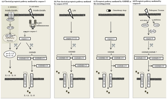
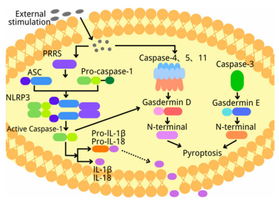
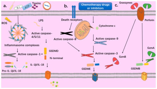
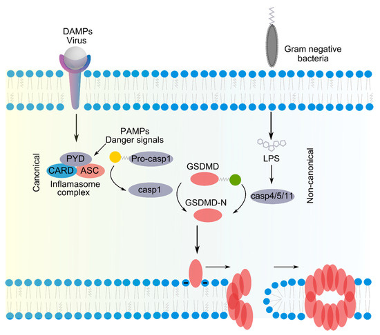
Fatty liver disease (FLD) is a clinical and pathological syndrome characterized by excessive fat deposition and even steatosis in hepatocytes. It has been proven that liver inflammation induced by fat and its derivatives are involved in the pathogenesis of FLD, while the precise mechanism still remains poorly understood. Pyroptosis is programmed inflammatory cell death driving cell swelling and membrane rupture. Pyroptosis is initiated by the activation of inflammasomes and caspases, which further cleaves and activates various gasdermins, leading to pores forming on the cell membrane and the release of pro-inflammatory factors such as interleukin (IL)-1β and IL-18. Recent studies demonstrate that pyroptosis occurs in hepatocytes, and inhibiting pyroptosis could effectively reduce fat deposition in the liver and could ameliorate inflammation from FLD, attracting our prime focus on the role of pyroptosis in FLD. In this manuscript, we reviewed the current understanding of pyroptosis in FLD development, aiming to provide new insights and potential research targets for the clinical diagnosis and intervention of FLD.
Full article
(This article belongs to the Section Molecular Pathology, Diagnostics, and Therapeutics)
►
Show Figures

Figure 1


