Int. J. Mol. Sci. 2023, 24(19), 14843; https://doi.org/10.3390/ijms241914843 - 2 Oct 2023
Cited by 1 | Viewed by 1155
Abstract
The severity of non-alcoholic fatty liver disease (NAFLD) ranges from simple steatosis to steatohepatitis, and it is not yet clearly understood which patients will progress to liver fibrosis or cirrhosis. SPARC (Secreted Protein Acidic and Rich in Cysteine) has been involved in NAFLD
[...] Read more.
The severity of non-alcoholic fatty liver disease (NAFLD) ranges from simple steatosis to steatohepatitis, and it is not yet clearly understood which patients will progress to liver fibrosis or cirrhosis. SPARC (Secreted Protein Acidic and Rich in Cysteine) has been involved in NAFLD pathogenesis in mice and humans. The aim of this study was to investigate the role of SPARC in inflammasome activation, and to evaluate the relationship between the hepatic expression of inflammasome genes and the biochemical and histological characteristics of NAFLD in obese patients. In vitro studies were conducted in a macrophage cell line and primary hepatocyte cultures to assess the effect of SPARC on inflammasome. A NAFLD model was established in SPARC knockout (SPARC−/−) and SPARC+/+ mice to explore inflammasome activation. A hepatic RNAseq database from NAFLD patients was analyzed to identify genes associated with SPARC expression. The results were validated in a prospective cohort of 59 morbidly obese patients with NAFLD undergoing bariatric surgery. Our results reveal that SPARC alone or in combination with saturated fatty acids promoted IL-1β expression in cell cultures. SPARC−/− mice had reduced hepatic inflammasome activation during the progression of NAFLD. NAFLD patients showed increased expression of SPARC, NLRP3, CASP1, and IL-1β. Gene ontology analysis revealed that genes positively correlated with SPARC are linked to inflammasome-related pathways during the progression of the disease, enabling the differentiation of patients between steatosis and steatohepatitis. In conclusion, SPARC may play a role in hepatic inflammasome activation in NAFLD.
Full article
(This article belongs to the Special Issue Molecular Advances in Liver Inflammation and Fibrosis)
►
Show Figures
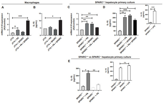
Figure 1

Figure 3

Figure 4

Figure 5

Figure 6


