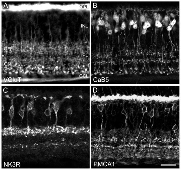
Fig. 1.
Fluorescence photomicrographs of vertical frozen sections through mouse retina that were labeled immunocytochemically. The retinal layers are indicated. OPL, outer plexiform layer; INL, inner nuclear layer; IPL, inner plexiform layer; GCL, ganglion cell layer. A: Conventional photomicrograph showing immunolabeling for the vesicular glutamate transporter 1 (VGluT1). Photoreceptor terminals in the OPL and bipolar cell axon terminals in the IPL are labeled. In the outer half of the IPL, putative OFF-cone bipolar cells terminate; in the inner half of the IPL, putative ON-cone bipolar cells and rod bipolar cells terminate. B: Confocal photomicrograph showing immunolabeling for the calcium-binding protein CaB5. Dendrites of bipolar cells in the OPL, their perikarya in the INL, and their axons terminating in three bands in the IPL are labeled. C: Confocal micrograph of a section immunolabeled for the neurokinin-3 receptor NK3R. Bipolar cells and their axons terminating in the outer IPL are labeled. The processes labeled in the inner IPL are amacrine cell dendrites. D: Confocal micrograph showing immunolabeling for the plasma membrane calcuim ATPase 1 (PMCA1). Photoreceptor terminals in the OPL, bipolar cell bodies in the INL, and bipolar cell axon terminals in the IPL are labeled. Scale bar = 19 μm for A, 20 μm for B,D, 17.7 μm for C.

