The
RYK gene encodes a receptor-like tyrosine kinase crucial for several biological processes, including development, tissue homeostasis, and cancer. This study utilized data from the Cancer Genome Atlas Project (TCGA) to evaluate
RYK expression at both mRNA and protein levels in various cancers,
[...] Read more.
The
RYK gene encodes a receptor-like tyrosine kinase crucial for several biological processes, including development, tissue homeostasis, and cancer. This study utilized data from the Cancer Genome Atlas Project (TCGA) to evaluate
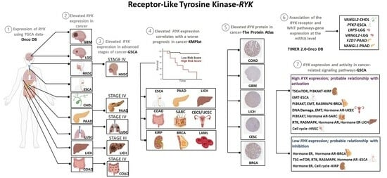
RYK expression at both mRNA and protein levels in various cancers, determine its prognostic significance, and explore its involvement in cancer-related signaling pathways. Elevated levels of
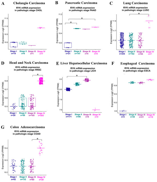
RYK mRNA were identified in cholangiocarcinoma (CHOL), pancreatic adenocarcinoma (PAAD), glioblastoma multiforme (GBM), lung squamous cell carcinoma (LUSC), brain lower grade glioma (LGG), head and neck squamous cell carcinoma (HNSC), liver hepatocellular carcinoma (LICH), esophageal carcinoma (ESCA), and colon adenocarcinoma (COAD), while
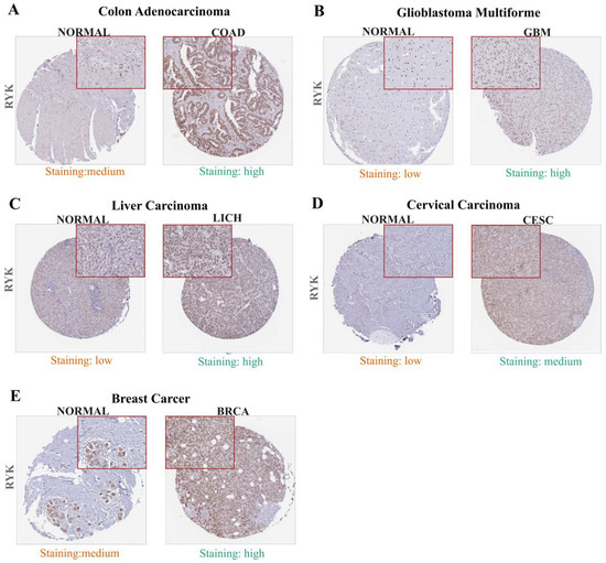
RYK protein levels were observed to be increased in colon adenocarcinoma (COAD), GBM, LICH, cervical and endocervical adenocarcinoma (CESC), and breast invasive carcinoma (BRCA). Additionally,
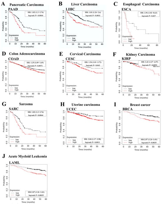
RYK overexpression correlated with poorer prognosis in several cancers, including PAAD, LICH, BRCA, ESCA, COAD, and CESC. Furthermore,
RYK showed a positive correlation with the upregulation of multiple receptors and coreceptors in the WNT signaling pathway in various types of cancer. In terms of cancer-related signaling pathways,
RYK was found to potentially interact with DNA damage, TSC/mTOR, PI3K/AKT, EMT, RTK, RAS/MAPK, ER hormone, AR hormone, and the cell cycle. This study provides new and previously unreported insights into the role of
RYK in cancer biology.
Full article


