Abstract
Deregulated expression of both Myc and Bcl-XL are consistent features of human plasma cell neoplasms (PCNs). To investigate whether targeted expression of Myc and Bcl-XL in mouse plasma cells might lead to an improved model of human PCN, we generated Myc transgenics by inserting a single-copy histidine-tagged mouse Myc gene, MycHis, into the mouse Ig heavy-chain Cα locus. We also generated Bcl-XL transgenic mice that contain a multicopy Flag-tagged mouse Bcl-xFlag transgene driven by the mouse Ig κ light-chain 3′ enhancer. Single-transgenic Bcl-XL mice remained tumor free by 380 days of age, whereas single-transgenic Myc mice developed B cell tumors infrequently (4 of 43, 9.3%). In contrast, double-transgenic Myc/Bcl-XL mice developed plasma cell tumors with short onset (135 days on average) and full penetrance (100% tumor incidence). These tumors produced monoclonal Ig, infiltrated the bone marrow, and contained elevated amounts of MycHis and Bcl-XLFlag proteins compared with the plasma cells that accumulated in large numbers in young tumor-free Myc/Bcl-XL mice. Our findings demonstrate that the enforced expression of Myc and Bcl-XL by Ig enhancers with peak activity in plasma cells generates a mouse model of human PCN that recapitulates some features of human multiple myeloma.
Introduction
Plasma cell neoplasms (PCNs) in humans comprise multiple myeloma (MM), Ig deposition and heavy-chain diseases, and plasmacytoma (PCT), which occurs as solitary PCT of bone and extramedullary PCT. Several mouse models of human PCN have been developed to study mechanisms of neoplastic plasma cell development and design new strategies for tumor treatment and prevention. Established mouse models of human PCN include tumors that arose spontaneously in old C57BL/KaLwRij mice and that resemble human MM (reviewed in ref. 1), peritoneal PCT that can be induced in strain BALB/c by intraperitoneal injections of proinflammatory agents and further accelerated by infection of mice with transforming retroviruses (reviewed in ref. 2), and the transplantation of human MM cells into SCID mice that harbor preimplanted human fetal bone as a nesting ground for the tumor cells (3–5). Currently emerging mouse models of human PCN are based on transgenic expression in B cells of IL-6 (6) and NPM-ALK (7) (fusion protein of nucleophosmin and anaplastic lymphoma kinase), or viral transduction of NPM-ALK in bone marrow cells (8). While all of these models afford valuable insights into the biology of human PCN, many important features of human PCN have not been adequately recapitulated in mice. One such feature with profound implications for pathogenesis, treatment, and prevention of human PCN is the collaboration of deregulated Myc (c-Myc) with tumor suppressor genes of the Bcl-2 family.
Deregulated expression of Myc is a consistent feature of PCN in humans and mice. In human MM, Myc appears to be activated in trans by a variety of signaling pathways converging at the Myc promoter (9, 10), possibly including IL-6 via Stat3 (11). Increased translation of Myc mRNA due to mutations in Myc’s internal ribosome entry site (12), stabilization of Myc protein via Ras (13) and other signaling pathways (14, 15), and chromosomal translocations deregulating Myc (16–18) may further contribute to Myc overexpression in MM. Upregulation of Myc may be of prognostic significance for MM patients (10). In contrast to human MM, the mechanism of Myc activation in BALB/c mouse PCT is uniform and well defined. Virtually all of these tumors harbor chromosomal translocations (19, 20) that activate Myc (21) by joining the Myc-Pvt1 locus at 15D1 with the Ig heavy-chain locus Igh at 12F2 or one of the Ig light-chain loci, Igκ at 6C1 or Igλ at 16A3, resulting in T(12;15)(Igh-Myc), T(6;15)(Igκ-Pvt1), and T(15;16)(Pvt1-Igλ), respectively. The most common translocation (∼90%) is T(12;15), which juxtaposes Myc in approximately 85% of T(12;15)-harboring tumors to the most downstream Igh gene, Cα. Mimicking the Myc-Igh(Cα) juxtaposition by gene insertion in mice might result in a mode of Myc deregulation that is conducive to plasmacytomagenesis and modeling of human PCN in mice.
Upregulation of death suppressor genes of the Bcl-2 family is another consistent feature of human and mouse PCN. Human MMs exhibit low levels of Bcl-2, but high levels of Mcl-1 and Bcl-XL (22), the main survival factors in MM (23–26). Overexpression of Bcl-XL via Stat3, a possible prognostic factor in MM (27), may be involved in growth factor independence of MM. In mouse PCT (28) and normal plasma cells in mice (29, 30), Bcl-XL, Bcl-2, and A1, rather than Mcl-1, are the main survival genes. The critical role of Bcl-XL in survival control of human MM and mouse PCT suggests that the enforced expression of this Bcl-2 family member is particularly promising for modeling of human PCN in mice. Studies on the biology of Bcl-XL and insights from transgenic mice expressing Bcl-XL (31), Bcl-2 (32, 33), Mcl-1 (34), or A1 (35) in B cells support this proposition. In mature B cells, Bcl-XL is upregulated in response to signaling through the B cell receptor (36–38), CD40 (39), and BAFF (Blys) (40). Bcl-XL enhances the survival of follicular and germinal center B cells (41), the presumed targets of the misguided DNA double-strand-break repair that generates chromosomal translocations including those involving Myc (42) in B cells undergoing V(D)J hypermutation (43) and isotype switching (44). Bcl-XL attenuates many death signals, resulting in the rescue of B cells that would normally be eliminated because of aberrant Ig gene rearrangements (31), autoreactivity (45), impaired affinity maturation (46), and genetic defects (47–50). Bcl-XL also mitigates Myc-dependent apoptosis in B cells, an important mechanism of Myc-induced lymphomagenesis (51). Targeting Bcl-XL expression to mature B and plasma cells might thus promote the neoplastic transformation of plasma cells, specifically those harboring Myc-activating chromosomal translocations or genetically engineered mutations that mimic such translocations in the germ line.
To test the possibility that deregulation of Myc and Bcl-XL in plasma cells might result in plasma cell tumors in mice, we developed two transgenic mouse strains. The first strain harbors a histidine-tagged mouse Myc gene, MycHis, inserted into the Cα gene of an otherwise intact mouse Igh locus that contains E∝, Eα, and all other regulatory elements residing in the Igh (Myc transgenics). The inserted Myc mimics the Myc-activating chromosomal T(12;15) translocation most commonly observed in peritoneal PCT of BALB/c mice. The second strain contains a multicopy Flag-tagged mouse Bcl-xFlag transgene driven by the mouse Ig κ light-chain 3′ enhancer (Bcl-XL transgenics). This transgene effects constitutive expression of Bcl-XL, the major splice form of Bcl-x mRNA, in B cells and plasma cells. Here we show that single-transgenic Myc and Bcl-XL mice exhibit moderate phenotypes with little or no impact on tumor development and lifespan of mice. In sharp contrast, double-transgenic Myc/Bcl-XL mice develop plasma cell tumors rapidly (135 days mean onset) and with full penetrance (100% tumor incidence). Our results show that novel targeted deregulation of Myc and Bcl-XL leads to a mouse model of human PCN that may be useful to elucidate the mechanism of the Myc/Bcl-XL collaboration and to design new approaches for treatment and prevention of human PCN.
Methods
Generation of Myc transgenics.
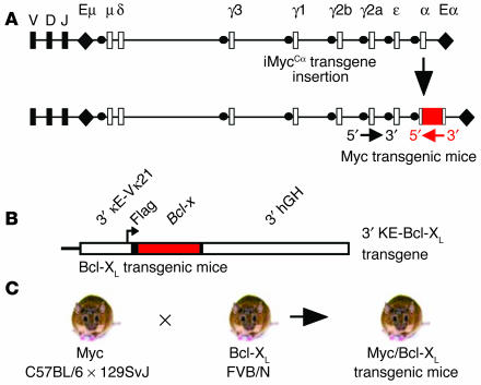
Gene targeting (52) and cre-loxP recombination (53) were used to insert a mouse Myc gene into the mouse germ-line Igh locus (Figure 1A). The inserted Myc consisted of an intron-less cDNA clone, the noncoding first exon with the natural P1/P2 promoter, 1.5 kb of genomic 5′ flank containing the normal transcription-regulatory region, and a short stretch of 3′ untranslated region (UTR) harboring the Myc major polyadenylation site. The UTR 3′ of the polyadenylation signal, which has been shown to be dispensable in vivo (54), was not present in the construct. The Myc coding region also contained an artificial histidine tag added in frame at its 3′ end. The tag made it possible to distinguish Myc mRNA and Myc protein encoded by the inserted MycHis gene from message and protein encoded by the normal, endogenous Myc gene. MycHis was inserted in opposite transcriptional orientation in intron 1 of the Cα locus. The construction of the inserted Myc, referred to as iMycCα, the assembly of the targeting vector, and the generation of the transgenic mice are described in the Supplemental Methods section and illustrated in Supplemental Figure 1 (suplemental material available at http://www.jci.org/cgi/content/full/113/12/1763/DC1). The Myc transgenic mice were of mixed C57BL/6 ∞ 129SvJ background (Figure 1C).
Figure 1.
Experimental overview of the generation of double-transgenic Myc/Bcl-XL mice. (A) Generation of Myc transgenic mice. Shown are the normal mouse Igh locus (top) and the targeted Igh locus with the inserted MycHis gene (bottom). The transcriptional orientation of Igh and MycHis is indicated by a black and a red arrow, respectively. (B) Generation of Bcl-XL transgenic mice. Depicted is a scheme of the Bcl-x transgene, which consists of the mouse 3′ κ enhancer; the promoter of the mouse variable κ gene, Vκ21; the mouse Bcl-x cDNA fused to the Flag epitope_encoding sequence; and the 3′ untranslated region of the human growth hormone (3′ hGH), a facilitator of Bcl-x expression. (C) Myc/Bcl-XL bitransgenics were on a mixed genetic background containing alleles from strains C57BL/6, 129SvJ, and FVB/N.
Generation of Bcl-XL transgenics.
Injection of plasmid DNA into male pronuclei of FVB/N zygotes was used to generate the Bcl-XL transgenic mice (Figure 1B). The transgene, referred to as 3′KE-Bcl-XL, was a modification of a previously developed Bcl-x transgene developed by Tim Behrens (University of Minnesota, Minneapolis, Minnesota, USA) (31). It contains the same Bcl-xFlag gene but uses the 3′ κ enhancer and VK21 promoter (excised from plasmid K3′E.KP.LUC; ref. 55) in place of the intronic E∝ enhancer and TK promoter to drive Bcl-x expression. Prior to microinjection, plasmid 3′κE-Bcl-xL was purified by cesium chloride centrifugation and linearized by digestion with NotI/AseI. Data presented in this report were collected using animals from the founder line 1967 (56). The Bcl-XL transgenic mice were of inbred FVB/N background (Figure 1C).
Generation of double-transgenic Myc/Bcl-XL mice.
Hemizygous Myc transgenic mice were crossed with hemizygous Bcl-XL transgenic mice to generate bitransgenics, which were born at the expected frequency of approximately 25%. The bitransgenic mice, henceforth referred to as Myc/Bcl-XL mice, were of mixed C57BL/6 ∞ 129SvJ ∞ FVB/N background (Figure 1C). Single-transgenic Myc and Bcl-XL F1 progeny (∼50% of offspring in the above-mentioned cross) and nontransgenic F1 progeny (∼25% of offspring) were used as controls unless otherwise noted. Breeding and maintenance of mice and all experimentation involving mice were approved under Institutional Animal Care and Use Committee Protocol 0006A56361 (University of Minnesota) and Animal Study Protocol LG-028 (National Cancer Institute [NCI]).
Histology and immunohistochemistry.
Tissues were fixed overnight in 10% buffered formalin and embedded in paraffin. Deparaffinized tissue sections were stained with H&E for histological examination by light microscopy or left unstained for marker analysis by immunohistochemistry. The latter involved incubation with biotin-conjugated antibodies to mouse B220 (RA3-6B2), CD138 (281-2; both from BD Biosciences, San Jose, California, USA), and Igκ (Southern Biotechnology Associates Inc., Birmingham, Alabama, USA), or incubation with unlabeled antibodies from rabbit to mouse phosphohistone H3 (Ser10) and cleaved caspase-3 (Asp175; both from Cell Signaling Technology Inc., Beverly, Massachusetts, USA) followed by a labeled secondary, goat anti-rabbit antibody. Antibody binding in tissue sections was visualized by the VECTASTAIN ABC alkaline phosphatase kit or the ABC peroxidase kit (Vector Laboratories Inc., Burlingame, California, USA) using Vector Blue or NovaRED as substrates. Endogenous alkaline phosphatase and peroxidase activities were inhibited by levamisole and hydrogen peroxide, respectively. Tissue sections were counterstained with hematoxylin.
ELISA.
Serum IgG and IgM concentrations were measured by ELISA using capture antibodies binding to both heavy chains and light chains (2 ∝g/ml; Caltag Laboratories Inc., Burlingame, California, USA). BSA (1%) was used for blocking. Serial dilutions of serum samples (1:2,000, 1:10,000, 1:50,000, 1:250,000, 1:500,000) were incubated at 4°C overnight in coated ELISA plates. Individual isotypes (IgG1, IgG2a, IgG2b, IgG3, and IgM) were detected with the help of biotinylated goat anti-mouse antibodies (Caltag Laboratories Inc.), followed by binding to alkaline phosphatase–conjugated streptavidin (Caltag Laboratories Inc.) and addition of 1 mg/ml alkaline phosphatase substrate in diethanolamine buffer. The optical densities were measured at 405 nm using a microplate reader.
Flow cytometry and cell sorting.
To determine subpopulations of lymphocytes in mouse tissues, single-cell suspensions from bone marrow, spleen, lymph nodes, and tumors were treated with FcBlock (anti–CD16/CD32, 2.4G2) and directly stained with mAb’s to mouse B220 (RA3-6B2), CD138 (281–2), CD3 (17A2), or IgM (II/41; all from BD Biosciences). These antibodies were conjugated to FITC, phycoerythrin (PE), allophycocyanin, or biotin. Isotype-specific controls demonstrated the specificity of labeling. To estimate the proliferation of normal B and plasma cells from spleen and bone marrow as well neoplastic plasma cells from plasma cell tumors, BrdU incorporation was measured in vitro according to the manufacturer’s protocol (BD Biosciences). Forty-eight hours after intraperitoneal injection of 0.5 mg BrdU, the cells were stained with allophycocyanin-labeled anti-B220 and PE-labeled anti-CD138 antibodies, fixed, permeabilized, treated with DNase, and incubated with a FITC-labeled antibody to BrdU. To analyze cell cycling in B and plasma cells, the cells were stained with 50 ∝g/ml propidium iodide (PI) in the presence of 0.1% sodium citrate and 0.1% Triton X-100. To determine rates of programmed cell death in B cells and plasma cells, the FITC-conjugated monoclonal active caspase-3 antibody apoptosis kit I from BD Biosciences was used (catalog number 550480). In all experiments, cells were analyzed on a Beckman Coulter FC 500 and sorted on an EPICS ALTRA (Beckman Coulter Inc., Fullerton, California, USA).
Cell sorting with magnetic beads (MACS).
To determine proliferation and apoptosis in B splenocytes, the MACS mouse B cell sorting kit with B220 beads (Miltenyi Biotec Inc., Auburn, California, USA) was used to purify B220+ cells. Briefly, spleen cells freed of red blood cells were separated on MACS VS+ columns using magnetic beads conjugated to an antibody to mouse B220 (CD45R). Recovery of B cells from single-cell suspensions was approximately 35%. The remaining cells were either B220– (∼35%) or lost (∼30%) during cell separation. The purity of MACS-sorted B220+ splenocytes was greater than 90%, as determined by FACS analysis.
Immunoblotting.
Proteins from clarified lysates of FACS-sorted homogenized cells from spleen, bone marrow, and plasma cell tumors were resolved electrophoretically in denaturing 10% SDS-PAGE gels and transferred by electroblotting to nitrocellulose membranes. Membranes were probed with rabbit anti-mouse antibodies to histidine tag (Cell Signaling Technology Inc.) and Flag tag (Sigma-Aldrich, St. Louis, Missouri, USA), or rabbit anti-mouse antibodies to cyclin D2 (sc-593), cyclin D1 (sc-717), and XBP-1 (sc-7160) from Santa Cruz Biotechnology Inc. (Santa Cruz, California, USA). The positions of the MycHis and Bcl-xLFlag proteins were visualized using the Phototope-HRP detection system (Cell Signaling Technology Inc.). To confirm equal loading, the membranes were stripped and reprobed using an antibody specific for actin (CLONTECH Laboratories Inc., Palo Alto, California, USA).
Southern analysis.
Genomic DNA (20 ∝g) was digested with BamHI and EcoRI, fractionated on a 0.7% agarose gel, transferred to a nylon membrane, and crosslinked under UV light. Following prehybridization (Hybrisol I; Intergen, Gaithersburg, Maryland, USA) at 42°C, the membrane was hybridized to a 1.1-kb Cκ probe labeled with 32P-CTP using a random-priming kit. The probe was generated by PCR using a primer pair that was designed by Michael Kuehl (NCI, NIH): 5′-GATGCTGCACCAACTGTATCCA-3′ and 5′-GGGGTGATCAGCTCTCAG-CTT-3′.
Paraproteins.
Paraproteins (M-spikes, extragradients) were detected using the Paragon SPE electrophoresis kit (Beckman Coulter Inc.). Ig isotypes were determined by ELISA using Immulon 2 plates (DYNEX Technologies Inc., Chantilly, Virginia, USA) and isotype-specific goat anti-mouse serum labeled with HRP (Southern Biotechnology Associates Inc.). Mouse serum samples were diluted from 10–3 to 1.28 ∞ 10–5. Plates were read on a Molecular Dynamics (Sunnyvale, California, USA) microplate reader at 450 nm.
DNA sequencing of 3′ VDJ regions.
The DNA sequence downstream of rearranged variable (V), diversity (D), and joining (J) gene segments of the Igh locus was determined as described elsewhere (57). DNA was amplified by PCR using a 5′ primer for the third framework region of VHJ558 genes and a 3′ primer that annealed 400 nucleotides downstream of the JH4 gene segment. The sequences of the forward and reverse primers used for tumor clones were 5′-AGCCTG-ACATCTGAGGAC-3′ and 5′-TAGTGTGGAACATTCCTCAC-3′, respectively. PCR amplification conditions were 95°C for 0.5 minutes, 63°C for 0.5 minutes, and 72°C for 1.5 minutes for 35 cycles. The sequences of the forward and reverse primers used for plasmablast clones (controls) were 5′-TTTGAATTCCTGACATCTGAGGACTCTGC-3′ and 5′-TTTGG-ATCCCTCCACCAGACCTCTCTAGA-3′, respectively. PCR conditions were 95°C for 0.5 minutes, 60°C for 0.5 minutes, and 72°C for 1.5 minutes for 30 cycles. PCR products from tumors were sequenced directly, and PCR products from controls were sequenced after cloning into pBluescript (Stratagene, La Jolla, California, USA) at the DNA sequencing facility of Iowa State University (Ames, Iowa, USA). An internal JH4 sequencing primer, iJH4 (5′-CTCCACCAGACCTCTCTAGA-3′), was used in either case.
Results
Features of single-transgenic Myc and Bcl-XL mice.
Myc transgenic mice were generated to recreate and study the molecular and cellular consequences of the Myc-activating T(12;15) translocations that characterize BALB/c PCT (Figure 1A). Several noteworthy features of the newly developed mouse strain suggest that insertion of MycHis into Igh resulted in representative modeling of T(12;15). First is the precise reconstruction of the translocation breakpoint region on mouse der(12); i.e., the 5′-to-5′ juxtaposition of MycHis and Cα. Second is the potential for MycHis to interact with the complete set of correctly spaced Eα enhancers, designed to create the same complex interplay of promoter and enhancer interactions that govern the expression of translocated Myc in mouse PCT. Third is the insertion of MycHis in the Igh chromatin domain, which presumably subjects the gene to the same higher-order regulatory influences (e.g., those imposed by chromatin remodeling and positional effects in the interphase nucleus) that affect Myc involved in the T(12;15) exchange. Bcl-XL mice were generated as part of a program to elucidate in transgenic mice the oncogenic properties of death suppressor genes that are frequently overexpressed in human MM (Figure 1B). The Bcl-x transgene is a modification of a previously developed Bcl-x transgene (31) in which the intronic heavy-chain enhancer E∝ was substituted with the 3′ κ enhancer (56). Compared with E∝, the 3′ κ enhancer delays Bcl-XLFlag expression during B cell development to the mature B cell and plasma cell stage (58), possibly favoring plasma cell tumor formation over B lymphoma development.
Plasmacytosis and hypergammaglobulinemia in double-transgenic Myc/Bcl-XL mice.
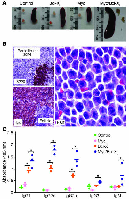
Enforced expression of Myc and Bcl-x by transcriptional control elements with peak activity in plasma cells (3′ Cα and 3′ κ enhancers) might result in expansion of plasma cells in vivo (plasmacytosis). To evaluate this possibility, four 8-week-old Myc/Bcl-XL mice (Figure 1C) were necropsied, and then lymphoid tissues were examined histologically and immunohistochemically. Four age-matched Myc transgenic mice, Bcl-XL transgenic mice, and nontransgenic littermates were used as controls. Myc/Bcl-XL transgenic mice invariably exhibited splenomegaly (Figure 2A), which was mainly caused by the massive accumulation of plasma cells in extrafollicular areas (Figure 2B). Immunostaining of tissue sections for B220 and κ light-chain expression, analogous to the sections shown in Figure 2B, demonstrated that the plasmacytosis sometimes involved nonlymphoid tissues, such as liver (Supplemental Figure 2). The increased tissue load with plasma cells was accompanied by a marked elevation of serum IgM and IgG in the Myc/Bcl-XL transgenic mice (Figure 2C). Bcl-XL transgenic mice also exhibited increases in serum Ig, but these were more moderate than in Myc/Bcl-XL transgenic mice and limited to three IgG isotypes (IgG3 was not higher in Bcl-XL mice than in Myc and normal mice). Myc transgenic mice were indistinguishable from normal mice (Figure 2C). ELISPOT analysis confirmed the ELISA results. It demonstrated a considerable increase in the number of antibody-producing cells in spleen and bone marrow of Myc/Bcl-XL transgenic mice compared with single-transgenic Myc and Bcl-XL mice and normal mice (Supplemental Figure 3a). Plasma cells obtained from tissues with plasmacytosis were not transplantable into pristane-primed BALB/c nude mice, suggesting that these cells had not yet undergone malignant transformation (Supplemental Figure 3b).
Figure 2.
Plasmacytosis and hypergammaglobulinemia in tumor-free Myc/Bcl-XL transgenic mice. (A) Splenomegaly in double-transgenic Myc/Bcl-XL mice relative to age-matched single-transgenic Myc and Bcl-XL mice and nontransgenic littermate controls. (B) Massive accumulation of plasmablasts and plasma cells in extrafollicular areas of the spleen in Myc/Bcl-XL transgenics. Left: Low-power view of follicular B cells (top) and plasma cells immunostained for B220 and κ light-chain expression, respectively (original magnification, ∞4). Right: High-power view of plasmablasts and plasma cells stained with H&E (original magnification, ∞63). (C) Marked elevation of serum Ig’s in Myc/Bcl-XL transgenic mice, measured by ELISA. *Significant difference (P < 0.05) by Student’s t test.
Figure 3.
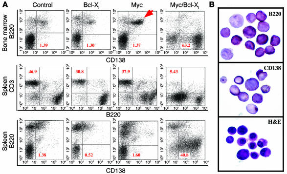
Plasmacytosis in Myc/Bcl-XL transgenic mice. (A) The percentage of B220–CD138+ plasma cells in the bone marrow (top row) and the spleen (bottom row) is indicated in the lower right quadrants of the depicted FACS scatter plots. The percentage of B220–CD3+ T cells in the spleen (center row) is indicated in the upper left quadrants. The red arrow denotes a population of B220+CD138+ plasmablasts in the bone marrow of Myc transgenic mice. (B) FACS-sorted B220+CD138+ cells from the bone marrow of Myc transgenic mice express B220 (top), CD138 (center), and Igκ (not shown) by immunostaining and exhibit cytological features of plasmablasts and plasma cells by H&E staining (bottom; original magnification, ∞63).
Bcl-XL promotes survival of MycHis-harboring plasmablasts.
To better quantify the apparent expansion of plasma cells in Myc/Bcl-XL transgenic mice, FACS analysis of bone marrow cells and splenocytes was performed. Six 6- to 10-week-old bitransgenics were compared with six age-matched single-transgenics and normal mice. Representative examples of each strain are shown in Figure 3A. Myc/Bcl-XL transgenic mice harbored up to 63% (average 42%, minimum 19%) B220–CD138hi plasma cells in the bone marrow, a striking increase compared with the Myc transgenics, Bcl-XL transgenics, and normal mice (Figure 3A, top). Similarly, Myc/Bcl-XL spleen contained up to 57% plasma cells (average 34%, minimum 14%), whereas spleen from the Myc transgenic, Bcl-XL transgenic, and normal mice contained less than 2% plasma cells (Figure 3A, bottom). Although the relative number of B220–CD3+ T cells was reduced in spleen from Myc/Bcl-XL bitransgenics compared with the other strains (Figure 3A, center), the absolute number of T cells remained comparable (because of the splenomegaly of strain Myc/Bcl-XL). Thus, neither transgene by itself, nor the combination of both transgenes grossly interfered with T cell homeostasis in the spleen. Studies with lymph nodes and bone marrow confirmed this observation (results not shown). A special feature of the Myc transgenic mice relative to all other mice was the presence of a distinct population of B220hiCD138+ cells in the bone marrow (22.5% in the example shown in the top panel of Figure 3A). Although the nature of these cells has not been fully elucidated, they likely represent plasmablasts (Figure 3B). The presence of expanded populations of plasmablasts in the Myc mice and plasma cells in the Myc/Bcl-XL mice suggested that most plasmablasts harboring only deregulated Myc fail to undergo terminal differentiation. However, when protected by Bcl-XL, Myc transgenic plasmablasts survive and mature into plasma cells.
Increased turnover of Myc-harboring B cells is reduced by Bcl-XL.
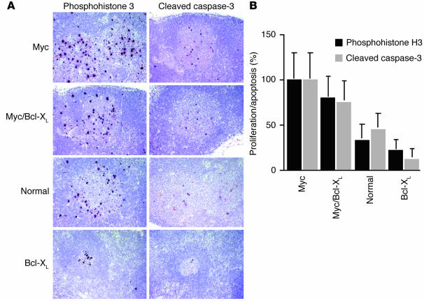
Myc’s potential to induce proliferation can be mitigated in vivo by Myc’s ability to trigger apoptosis (59). To compare proliferation and apoptosis in mature B cells of Myc/Bcl-XL bitransgenic mice with those in single-transgenic Myc and Bcl-XL mice and nontransgenic littermates, tissue sections of lymph nodes from 8-week-old mice were immunostained with antibody to phosphohistone H3 (a marker of mitosis) and cleaved caspase-3 (a marker of apoptosis). B cell proliferation in lymph nodes, predominantly in follicles and germinal centers, was most vigorous in Myc transgenic mice, followed by Myc/Bcl-XL transgenic, normal, and Bcl-XL transgenic mice, which had the lowest (Figure 4). To better evaluate the apparent increase in Myc-dependent proliferation, FACS analysis of PI-stained cells was combined with BrdU labeling in vivo. Myc-harboring B cells proliferated nearly 2.5 times faster by PI staining (Supplemental Figure 4a, left) and three times faster by PI/BrdU staining (Supplemental Figure 4a, center) than the controls. Apoptosis in B cells measured by immunostaining for cleaved caspase-3 was also highest in Myc transgenic mice, lowest in Bcl-XL transgenic mice, and intermediate in Myc/Bcl-XL and normal mice (Figure 4). TUNEL of lymph node, spleen, and bone marrow sections confirmed this result (not shown). To better assess the apparent increase in Myc-dependent apoptosis, freshly isolated Myc-harboring B cells were analyzed by FACS for activation of caspase-3. Apoptosis in these B cells was elevated approximately 4.5-fold compared with normal B cells (Supplemental Figure 4a, right). The propensity of Myc-containing B cells to undergo apoptosis was further increased (about twofold) upon activation of cells in vitro with LPS, anti-IgM, or both (Supplemental Figure 4b). Together, these findings indicated that the high turnover of B cells in the Myc transgenic mice is attenuated by Bcl-XL.
Figure 4.
Proliferation and apoptosis in lymph node follicles of 8-week-old Myc, Myc/Bcl-XL, and Bcl-XL mice compared with normal nontransgenic littermates. (A) Sections of a peripheral lymph node containing two follicles. Follicular B cells undergoing proliferation or apoptosis are immunostained (brown spots) for phosphohistone H3 or activated caspase-3, respectively (original magnification, ∞40). The images are ordered from top to bottom according to the apparent turnover of follicular B cells, which was highest in the Myc mice and lowest in the Bcl-XL mice. (B) The result of the microscopic enumeration of proliferating B cells (black bars) and dying B cells (gray bars) in lymph node follicles. Three lymph nodes of each mouse strain were evaluated to determine mean values and SDs of the mean.
Myc/Bcl-XL CD138+ cells actively proliferate.
Normal plasma cells are end-stage B cells that have lost the ability to proliferate. To evaluate whether plasma cells of Myc/Bcl-XL transgenic mice adhered to this rule, immunostaining of tissue sections for phosphorylated histone H3 was combined with immunostaining for syndecan-1 (CD138, a marker for plasmablasts and plasma cells) or B220 (CD45, a pan–B cell marker). Doubly stained spleen sections from 8-week-old mice clearly showed that CD138+ cells participated in cell cycling (Supplemental Figure 5a, left). The same result was obtained with lymph node and bone marrow sections (not shown), and when BrdU incorporation in vivo was used as the indicator of proliferation instead of phosphorylated histone H3 (not shown). Double staining of spleen sections (Supplemental Figure 5a, right) or lymph node and bone marrow sections (not shown) for cleaved caspase-3 and CD138 indicated that Myc/Bcl-XL plasmablasts and plasma cells underwent apoptosis. This observation was confirmed with TUNEL of lymph node, spleen, and bone marrow sections (data not shown). Of importance, the semiquantitative comparison of the extent of proliferation and apoptosis in serial tissue sections of double-transgenic mice, such as those shown in Supplemental Figure 5b, indicated that although there was ongoing apoptosis in Myc/Bcl-XL CD138+ cells, the balance was tipped in favor of proliferation. This resulted in an enormous expansion of plasma cells, which was unique to the Myc/Bcl-XL mice and not observed in single-transgenic and control mice. These findings suggested that the interaction of Myc and Bcl-XL results in an expanded pool of actively proliferating CD138+ cells (most likely plasmablasts) in the double-transgenic mice.
Figure 5.
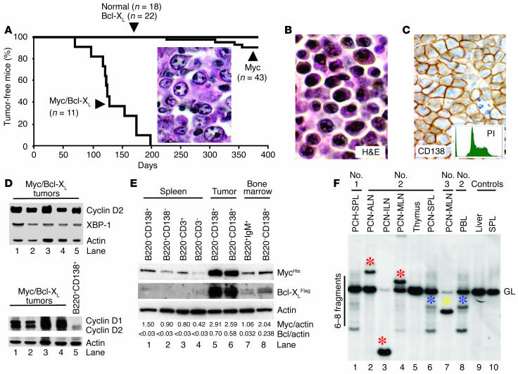
Plasma cell neoplasms in Myc/Bcl-XL transgenic mice. (A) Bcl-XL transgenic mice (n = 22), nontransgenic littermates (n = 10, not shown), and C57BL/6 mice (n = 18) remained tumor free. Three of four tumors that developed in Myc transgenic mice (n = 43) were diffuse large B cell lymphomas containing abundant neoplastic centroblasts (inset, H&E; original magnification, _63). (B) Typical Myc/Bcl-XL plasma cell tumor stained with H&E (original magnification, _63). (C) Myc/Bcl-XL plasma cell neoplasm immunostained for CD138 (brown; original magnification, _40). The inset shows a FACS profile of the vigorously proliferating propidium iodine_stained tumor cells. (D) Western analysis of five Myc/Bcl-XL plasma cell tumors for expression of cyclin D2 (Myc target), XBP-1 (plasma cell transcription factor), and actin (loading control, top panel). Cyclin D1/D2 expression in Myc/Bcl-XL plasma cell tumors (lanes 1_4) compared with “premalignant” plasmablasts from tumor-free Myc/Bcl-XL mice (lane 5, bottom panel). (E) Western analysis of MycHis and Bcl-XLFlag expression in flow-sorted Myc/Bcl-XL tumor cells (lanes 5_6) compared with cells from spleen (lanes 1_4) and bone marrow (lanes 7_8) of tumor-free Myc/Bcl-XL mice. Bcl-XL occurs in two alternative splice forms, with and without transmembrane domain. Myc and Bcl-XL were detected with antibodies against the histidine and Flag epitopes. (F) Southern analysis for clonotypic Igκ rearrangements in PCNs (lanes 2_4, 6, and 7), spleen (SPL) with plasma cell hyperplasia (PCH; lane 1), thymus (lane 5), and peripheral blood leukocytes (PBL; lane 8) from three Myc/Bcl-XL mice compared with liver and spleen from nontransgenic littermates. ALN, axillary LN; ILN, inguinal LN; MLN, mesenteric LN; GL, germline.
Rapid development of plasma cell tumors in Myc/Bcl-XL transgenic mice.
To evaluate whether the sustained proliferation of Myc/Bcl-XL plasma cells leads to the development of plasma cell neoplasms, 11 double-transgenic mice were monitored for tumor incidence and survival (Figure 5A). Unlike Bcl-XL transgenic mice (n = 22) and normal mice (n = 18), which remained tumor free by 380 days of age, Myc/Bcl-XL mice exhibited a drastically reduced survival. This was caused by malignant plasma cell tumors (Figure 5B) that developed rapidly (mean onset 135 days) and with full penetrance (incidence 100%). Myc transgenic mice (n = 43) also developed neoplasms, but tumor development took a long time (mean onset 330 days), occurred with low penetrance (incidence 9.3%), and resulted in B cell lymphomas rather than plasma cell tumors: diffuse large B cell lymphoma in three of four cases (Figure 5A, inset) and unclassified B lymphoma in one case (not shown). The weak tumor phenotype of the Myc mice suggested that although the MycHis transgene reproduced the requisite molecular changes that initiate neoplastic B cell and plasma cell development in mice, MycHis’s true oncogenic potential in vivo was tempered in the absence of Bcl-XL.
Features of Myc/Bcl-XL plasma cell tumors.
Plasma cell neoplasms of Myc/Bcl-XL transgenic mice were CD138+ by immunohistochemistry (Figure 5C), proliferated vigorously by flow cytometry of PI-stained tumor cells (Figure 5C, inset), and expressed transcription factors typical of plasma cells, such as XBP-1 and Blimp-1 message (RT-PCR results not shown) and XBP-1 protein (Figure 5D, top). Immunoblotting showed that Myc/Bcl-XL tumor cells expressed Myc and Bcl-XL at higher levels (Figure 5E, lanes 5 and 6) than plasma cells from tumor-free Myc/Bcl-XL mice (lanes 1 and 8). Flow-sorted tumor cells had a mean MycHis/actin ratio of 2.75 and thus contained 1.8-fold and 2.6-fold higher MycHis protein levels than flow-sorted plasma cells from tumor-free spleen (lane 1) and bone marrow (lane 8), respectively. Consistent with the high Myc level, cyclin D2, a validated Myc target (60), and cyclin D1 were overexpressed in the tumors (Figure 5D, bottom, lanes 1–4) relative to B220+CD138+ plasmablasts from tumor-free Myc/Bcl-XL mice (control, lane 5). Myc/Bcl-XL tumors were readily transplantable upon transfer of fewer than 106 tumor cells into pristane-primed BALB/c nude mice, with tumor take occurring in less than 2 weeks in two of two cases (not shown). Continuous cell lines were readily derived from two additional cases of primary plasma cell neoplasia. Together, these findings indicated that Myc/Bcl-XL transgenic mice are predisposed to plasma cell neoplasms that exhibit high levels of MycHis and Bcl-XLFlag and that have completed malignant transformation.
Origin and distribution of plasma cell tumors.
The presentation of Myc/Bcl-XL transgenic mice with plasma cell tumors in multiple tissues raised the question of whether the tumors originated from a single precursor with the potential for early, widespread dissemination (monocentric origin), or from multiple precursors resulting in the outgrowth of tumors with different molecular features (multicentric origin). To investigate this, genomic tumor DNA was analyzed by Southern hybridization for clonotypic Igκ rearrangements (Figure 5F). Three different states of tumor dissemination were observed, often coexisting in the same mouse. Some tissues, such as the spleen of mouse no. 1, a borderline case of plasma cell hyper- and neoplasia, harbored multiple clones of aberrant plasma cells (lane 1), reminiscent of the clonal diversity observed in early stages of peritoneal plasmacytomagenesis in BALB/c mice (61). Other tissues, such as the mesenteric lymph node of mouse no. 3 (lane 7), contained one dominant tumor clone (yellow asterisk) that had essentially replaced the normal tissue, as reflected by the loss of the κ germ-line fragment. A third category of tissues, e.g., the lymph nodes in mouse no. 2 (lanes 2–4), harbored different tumor cell clones with distinct κ rearrangements (red asterisks). The detection of an additional cell clone in the peripheral blood leukocyte sample of the same mouse (lane 8, blue asterisk) indicated the emergence of a leukemic clone that had infiltrated the spleen (lane 6) but had not yet reached the thymus (lane 5). These findings reflected a considerable variability in the progression of Myc/Bcl-XL plasma cell tumors. Some tumors remained confined to lymphoid tissues, where they evolved in mono- or multicentric fashion, whereas other tumors acquired the ability for systemic dissemination, leading to full-blown plasma cell leukemia in some cases (not shown).
Bone marrow involvement of plasma cell tumors.
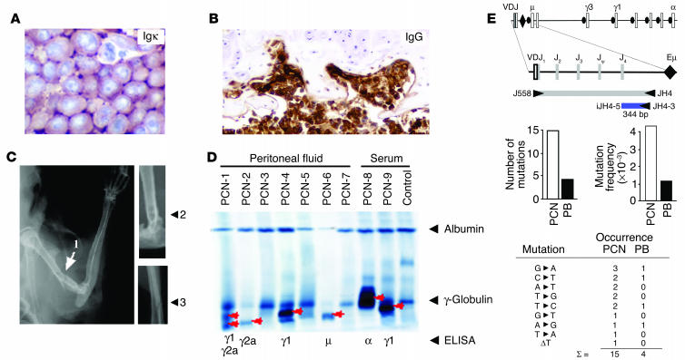
Myc/Bcl-XL plasma cell tumors demonstrated proclivity to bone marrow involvement, generating coalescent, wall-to-wall tumor masses in several mice with terminal neoplasia (Figure 6A). Histological examination of bone sections from mice with less advanced tumors routinely revealed multifocal lesions of aberrant, pleomorphic plasma cells adjacent to diminished or dissolved osseous trabeculae or located in resorption pits at the inner surface of the corticalis (Figure 6B). Some bone sections contained large sheets of neoplastic plasma cells in soft tissues surrounding the bones, indicating that the tumor had penetrated the corticalis at an obscure, nearby site. Whole-body radiographs showed osteolytic lesions and putative pathological fractures in long bones of three mice (Figure 6C). Serum and peritoneal lavage fluid of tumor-bearing mice often contained monoclonal Ig spikes (extragradients, M components) that were readily detectable by protein electrophoresis (Figure 6D). Two of three tumors from mice without evidence for M-spikes in lavage samples (Figure 6D, lanes 3, 5, and 7) did not express heavy chain (PCN-3) or light chain (PCN-7) by Western analysis (result not shown), possibly because Ig genes were deleted because of genomic instability in the tumors. To determine whether Myc/Bcl-XL plasma cell tumors exhibit evidence for hypermutation of expressed Ig genes, the 344-bp 3′ JH4 region just downstream of the rearranged VDJ gene was sequenced (Figure 6E, top). Ten tumor-derived clones with unique VH rearrangements were assessed for frequency and type of somatic mutations, and then compared with ten analogous clones from plasmablasts of tumor-free Myc/Bcl-XL mice (controls). Fifteen mutations were identified in the tumors, and four mutations were found in the plasmablasts (P = 0.022, χ2 analysis; Figure 6E, center left). The corresponding mutation frequencies in the tumors and plasmablasts were 4.36 ∞ 10–3 per bp and 1.16 ∞ 10–3 per bp, respectively (Figure 6E, center right). All mutations except one (a 1-bp deletion) were base substitutions (Figure 6E, bottom). Three mutations in the tumors occurred at WA dinucleotides (W = A or T), a preferred target of the VDJ hypermutation machinery (Supplemental Figure 6). Although these findings are too preliminary for firm conclusions, they suggest that Myc/Bcl-XL plasma cell tumors have germinal center experience.
Figure 6.
Myc/Bcl-XL PCNs infiltrate the bone marrow, produce monoclonal Ig, and cause osteolytic lesions. (A) Bone marrow infiltration with Igκ-producing neoplastic plasma cells (original magnification, _63). (B) Palisades of neoplastic plasma cells (brown) destroying the luminal face of a femur’s corticalis (IgG immunostaining; original magnification, _40). Two bone resorption lacunae are denoted by arrows. (C) Radiographs of large osteolytic lesion with apparent pathological fracture (1, left humerus), osteolytic lesion without fracture (2, right femur), and hairline fracture without visible osteolytic lesion (3, left forearm). (D) Protein electropherogram of serum and peritoneal lavage samples containing M-spikes (red arrows) isotyped using ELISA (bottom). (E) Frequency and type of mutations in the 3′ JH4 region of rearranged variable (V) genes. Shown at the top is a scheme of the mouse Igh locus. PCR primers J558 and JH4 were used to amplify rearranged VDJ genes and linked 3′JH4 sequences. Sequencing primers iJH4-5′ and iJH4-3′ were used to detect mutations in the 344-bp 3′ JH4 region. Shown in the center are bar diagrams of the number of mutations in the 3′ JH4 region in PCNs and plasmablasts from tumor-free Myc/Bcl-XL mice (PB, left panel). The corresponding mutation frequencies (mutations/3440 bp) are plotted to the right. Shown at the bottom are types and occurrences of base substitution mutations in the 3′ JH4 region of rearranged VH genes in PCN and PB. The tumor sample also contained a deletion, δT. The location of the mutation in the 3′ JH4 regions is depicted in Supplemental Figure 6.
Discussion
This study reports the development of a rapid-onset high-penetrance mouse model of human PCN that is based on forced coexpression of Myc and Bcl-XL in plasma cells. Novel targeted deregulation of Myc (insertion in the proximity of the heavy-chain 3′-Cα enhancer) and Bcl-x (activation by the κ light-chain 3′ enhancer) resulted in the formation of transplantable, Ig-producing, CD138+ plasma cell tumors in double-transgenic Myc/Bcl-XL mice. Tumor development was preceded by massive expansion of normal plasma cells (generalized plasmacytosis) admixed with atypical, dividing plasmablasts (plasma cell hyperplasia), the possible targets of the combined oncogenic attack of Myc and Bcl-XL. The striking tumor phenotype in Myc/Bcl-XL bitransgenics — relative to the weak tumor phenotype in single-transgenic Myc mice and the apparent absence of tumors in single-transgenic Bcl-XL mice — demonstrated that complementation of a proliferation-inducing oncogene (Myc) with a death suppressor gene (Bcl-x) can greatly accelerate plasma cell neoplasms in mice. This finding was consistent with previous studies on plasmacytomagenesis in mice and recent insights into the biology of Myc and Bcl-XL in B cells and plasma cells in humans and mice.
The weak oncogenic potency of MycHis, on its own, was somewhat surprising. It was probably caused by Myc-dependent apoptosis of tumor precursors, rather than failure of the transgenic construct to recreate the biological features of the Myc-activating T(12;15) translocation seen in BALB/c PCT. Studies on Myc and PCT development in BALB/c mice support this contention. Although, normally, Myc promotes cell growth and proliferation in the presence of growth factors (62, 63), deregulated Myc, in the absence of growth factors, can force quiescent cells into active cell cycle (64, 65) and then trigger apoptosis. Myc has been shown to augment apoptosis by suppressing Bcl-XL (66). Myc-induced apoptosis is a safeguard mechanism for eliminating aberrant cells with active Myc (67, 68). In agreement with this, PCT induction studies in BALB/c mice have shown that tumor precursors containing activated Myc are removed when positive survival signals provided by IL-6 (69, 70) and environmental antigen stimulation (71, 72) are missing or limiting. Likewise, PCR studies on the occurrence of T(12;15) translocations in mice have demonstrated that the majority of T(12;15)-harboring cells do not evolve into PCT (reviewed in ref. 73), presumably because they are eliminated by Myc-induced apoptosis. Although the principal target of Myc-dependent apoptosis during tumor development is not known, our observations in the Myc/Bcl-XL model suggest that the MycHis-harboring plasmablast is a candidate. Additional studies are warranted to elucidate Bcl-XL’s survival function in Myc-activated plasmablasts.
The finding that 11 of 11 Myc/Bcl-XL bitransgenics developed plasma cell tumors by 200 days of age demonstrated that Bcl-XL collaborates with Myc to promote neoplastic plasma cell development. Studies on lymphomagenesis in E∝-Myc transgenic mice (74) have provided intriguing insights concerning the underlying biology of the Myc/Bcl-XL collaboration. E∝-Myc–driven B cell tumors appear to depend on abrogation of Myc-induced apoptosis, e.g., by selection for mutants with upregulated expression of Bcl-2–family proteins, such as Bcl-XL (75). However, the fact that Bcl-XL attenuates apoptosis does not exclude that Bcl-XL uses additional mechanisms to accelerate PCT in mice. Bcl-XL can delay Myc-induced cell cycle entry by interfering with the ability of Myc to downregulate p27 and activate cyclin/cdk complexes (76). Bcl-XL can exert promutagenic and genome-destabilizing effects by reducing DNA repair efficiency (77), a possible factor in the development of inflammation-induced PCT in BALB/c mice (78–80). Bcl-XL is involved in bypassing growth factor requirements and resistance to anoikis (death initiated by loss of contact with ECM components) (81), which may be important for the mobilization and systemic dissemination of malignant plasma cells. Finally, as mentioned above, Bcl-XL may facilitate the terminal differentiation of MycHis plasmablasts, which would be in line with Bcl-XL’s differentiation-enhancing properties in B lymphocytes (45, 50). Thus, although the mechanism of the Myc/Bcl-XL collaboration may be complex, and although differences in Myc deregulation between E∝-Myc mice and MycHis mice may further modify this collaboration, the findings in the E∝-Myc model strongly suggest that protection from Myc-induced apoptosis is a critical component of Bcl-XL’s ability to promote plasma cell tumor formation in Myc/Bcl-XL mice.
Bone marrow infiltration with malignant plasma cells, a consistent feature of neoplasms arising in Myc/Bcl-XL mice, indicated that further modification of strain Myc/Bcl-XL might lead to an improved mouse model of human MM. Possible approaches for modeling human MM in Myc/Bcl-XL transgenic mice involve the transgenic expression of (a) MM progressor genes, such as ABL, FGFR3, RAS, and WNT, (b) transcription factors that commit B cells to the plasma cell fate, such as Blimp-1, IRF4, and XBP-1, (c) chemokine receptors that direct plasma cells from lymph node to bone marrow, such as CXCR4, (d) adhesion molecules that mediate the interaction between plasma cells and stroma cells and/or ECM in the bone marrow, such as syndecan-1, VLA-4, and osteoprotegerin, and (e) mediators of bone destruction, such as MIP-1α, RANKL, and DKK1. Considering that human MM occurs predominantly in elderly patients, it may also become important to delay the expression of the Myc and Bcl-x transgenes, because their inducible expression in aging mice and their constitutive expression in young mice may favor different types of plasma cell tumor. Furthermore, the well-established importance of modifier genes in peritoneal BALB/c PCT (82) suggests that it may also be necessary to introduce the Myc and Bcl-x transgenes on a genetic background (e.g., DBA/2N) that is resistant to peritoneal (extramedullary) PCT and, therefore, possibly more conducive to plasma cell tumor formation in the bone marrow.
In conclusion, the remarkable efficiency with which Bcl-XL synergizes with Myc in plasma cell tumor development in mice extends studies by other investigators who have used Bcl-XL to promote Myc-dependent oncogenesis in pancreas (59), skin, and other tissues (83). It further extends work with two independently developed Bcl-2 transgenes that facilitated the malignant transformation of B cells and plasma cells harboring deregulated Myc (84, 85). Plasma cell tumors in Myc/Bcl-XL transgenics may afford a good model system for studying the mechanism by which overexpression of Myc and Bcl-XL facilitates human PCN. It would be particularly interesting to elucidate whether the known mechanisms of Myc enhancement at the transcriptional level (NF-κB [ref. 86] and Stat3 [ref. 87]) and/or the protein level (Ras [ref. 13], NF-κB [ref. 14], and CK2 signaling [ref. 15]) are also operational in the Myc/Bcl-XL plasma cell tumor model. This information and other insights gleaned from Myc/Bcl-XL mice may lead to new interventions to inhibit the Myc/Bcl-XL collaboration for the benefit of the human PCN patient.
Supplementary Material
Acknowledgments
We wish to thank Lino Tessarollo and Eileen Southon (Gene Targeting Core, NCI) for generating iMycCα mice; Sung Sup Park (Korean Research Institute of Bioscience and Biotechnology, Taejon, Republic of Korea) for constructing an early version of the iMycCα gene targeting vector; the staff of the Mouse Genetics and Histopathology Cores, University of Minnesota, for generating and analyzing 3′KE-Bcl-XL mice; the staff of the mouse facility of the Laboratory of Genetics, Center for Cancer Research (NCI), particularly Wendy DuBois, Lisa Craig, and Vaishali Jarral, for help with the in vivo studies; Sandra Schieferl and Michael Lewis (Cell Signaling Technology Inc.) for outstanding technical assistance; and Michael Comb (Cell Signaling Technology Inc.) and Beverly Mock (NCI) for encouragement and scientific discussion. This study was supported in part by NCI Cancer Biology Training grant T32 CA09138-27 to M. Linden and a grant from the Leukemia Research Fund to B. Van Ness.
Footnotes
Wan Cheung Cheung and Joong Su Kim contributed equally to this work.
Nonstandard abbreviations used: multiple myeloma (MM); National Cancer Institute (NCI); plasma cell neoplasm (PCN); plasmacytoma (PCT).
Conflict of interest: The authors have declared that no conflict of interest exists.
References
- 1.Radl J. Multiple myeloma and related disorders. Lessons from an animal model. Pathol. Biol. (Paris). 1999;47:109–114. [PubMed] [Google Scholar]
- 2.Potter M. Experimental plasmacytomagenesis in mice. Hematol. Oncol. Clin. North Am. 1997;11:323–347. doi: 10.1016/s0889-8588(05)70434-2. [DOI] [PubMed] [Google Scholar]
- 3.Yaccoby S, Barlogie B, Epstein J. Primary myeloma cells growing in SCID-hu mice: a model for studying the biology and treatment of myeloma and its manifestations. Blood. 1998;92:2908–2913. [PubMed] [Google Scholar]
- 4.Yaccoby S, Epstein J. The proliferative potential of myeloma plasma cells manifest in the SCID-hu host. Blood. 1999;94:3576–3582. [PubMed] [Google Scholar]
- 5.Pearse RN, et al. Multiple myeloma disrupts the TRANCE/ osteoprotegerin cytokine axis to trigger bone destruction and promote tumor progression. Proc. Natl. Acad. Sci. U. S. A. 2001;98:11581–11586. doi: 10.1073/pnas.201394498. [DOI] [PMC free article] [PubMed] [Google Scholar]
- 6.Kovalchuk AL, et al. IL-6 transgenic mouse model for extraosseous plasmacytoma. Proc. Natl. Acad. Sci. U. S. A. 2002;99:1509–1514. doi: 10.1073/pnas.022643999. [DOI] [PMC free article] [PubMed] [Google Scholar]
- 7.Chiarle R, et al. NPM-ALK transgenic mice spontaneously develop T-cell lymphomas and plasma cell tumors. Blood. 2003;101:1919–1927. doi: 10.1182/blood-2002-05-1343. [DOI] [PubMed] [Google Scholar]
- 8.Lange K, et al. Overexpression of NPM-ALK induces different types of malignant lymphomas in IL-9 transgenic mice. Oncogene. 2003;22:517–527. doi: 10.1038/sj.onc.1206076. [DOI] [PubMed] [Google Scholar]
- 9.De Vos J, et al. Comparison of gene expression profiling between malignant and normal plasma cells with oligonucleotide arrays. Oncogene. 2002;21:6848–6857. doi: 10.1038/sj.onc.1205868. [DOI] [PubMed] [Google Scholar]
- 10.Zhan F, et al. Global gene expression profiling of multiple myeloma, monoclonal gammopathy of undetermined significance, and normal bone marrow plasma cells. Blood. 2002;99:1745–1757. doi: 10.1182/blood.v99.5.1745. [DOI] [PubMed] [Google Scholar]
- 11.Kiuchi N, et al. STAT3 is required for the gp130-mediated full activation of the c-myc gene. J. Exp. Med. 1999;189:63–73. doi: 10.1084/jem.189.1.63. [DOI] [PMC free article] [PubMed] [Google Scholar]
- 12.Chappell SA, et al. A mutation in the c-myc-IRES leads to enhanced internal ribosome entry in multiple myeloma: a novel mechanism of oncogene de-regulation. Oncogene. 2000;19:4437–4440. doi: 10.1038/sj.onc.1203791. [DOI] [PubMed] [Google Scholar]
- 13.Sears R, Leone G, DeGregori J, Nevins JR. Ras enhances Myc protein stability. Mol. Cell. 1999;3:169–179. doi: 10.1016/s1097-2765(00)80308-1. [DOI] [PubMed] [Google Scholar]
- 14.Grumont RJ, Strasser A, Gerondakis S. B cell growth is controlled by phosphatidylinosotol 3-kinase-dependent induction of Rel/NF-κB regulated c-myc transcription. Mol. Cell. 2002;10:1283–1294. doi: 10.1016/s1097-2765(02)00779-7. [DOI] [PubMed] [Google Scholar]
- 15.Channavajhala P, Seldin DC. Functional interaction of protein kinase CK2 and c-Myc in lymphomagenesis. Oncogene. 2002;21:5280–5288. doi: 10.1038/sj.onc.1205640. [DOI] [PubMed] [Google Scholar]
- 16.Shou Y, et al. Diverse karyotypic abnormalities of the c-myc locus associated with c-myc dysregulation and tumor progression in multiple myeloma. Proc. Natl. Acad. Sci. U. S. A. 2000;97:228–233. doi: 10.1073/pnas.97.1.228. [DOI] [PMC free article] [PubMed] [Google Scholar]
- 17.Avet-Loiseau H, et al. Rearrangements of the c-myc oncogene are present in 15% of primary human multiple myeloma tumors. Blood. 2001;98:3082–3086. doi: 10.1182/blood.v98.10.3082. [DOI] [PubMed] [Google Scholar]
- 18.Fabris S, et al. Heterogeneous pattern of chromosomal breakpoints involving the MYC locus in multiple myeloma. Genes Chromosomes Cancer. 2003;37:261–269. doi: 10.1002/gcc.10211. [DOI] [PubMed] [Google Scholar]
- 19.Ohno S, et al. Nonrandom chromosome changes involving the Ig gene-carrying chromosomes 12 and 6 in pristane-induced mouse plasmacytomas. Cell. 1979;18:1001–1007. doi: 10.1016/0092-8674(79)90212-5. [DOI] [PubMed] [Google Scholar]
- 20.Wiener F, et al. High resolution banding analysis of the involvement of strain BALB/c- and AKR-derived chromosomes No. 15 in plasmacytoma-specific translocations. J. Exp. Med. 1984;159:276–291. doi: 10.1084/jem.159.1.276. [DOI] [PMC free article] [PubMed] [Google Scholar]
- 21.Shen-Ong GL, Keath EJ, Piccoli SP, Cole MD. Novel myc oncogene RNA from abortive immunoglobulin-gene recombination in mouse plasmacytomas. Cell. 1982;31:443–452. doi: 10.1016/0092-8674(82)90137-4. [DOI] [PubMed] [Google Scholar]
- 22.Krajewski S, et al. Immunohistochemical analysis of in vivo patterns of Bcl-X expression. Cancer Res. 1994;54:5501–5507. [PubMed] [Google Scholar]
- 23.Zhang B, Gojo I, Fenton RG. Myeloid cell factor-1 is a critical survival factor for multiple myeloma. Blood. 2002;99:1885–1893. doi: 10.1182/blood.v99.6.1885. [DOI] [PubMed] [Google Scholar]
- 24.Spets H, et al. Expression of the bcl-2 family of pro- and anti-apoptotic genes in multiple myeloma and normal plasma cells: regulation during interleukin-6(IL-6)-induced growth and survival. Eur. J. Haematol. 2002;69:76–89. doi: 10.1034/j.1600-0609.2002.01549.x. [DOI] [PubMed] [Google Scholar]
- 25.Zhang B, Potyagaylo V, Fenton RG. IL-6-independent expression of Mcl-1 in human multiple myeloma. Oncogene. 2003;22:1848–1859. doi: 10.1038/sj.onc.1206358. [DOI] [PubMed] [Google Scholar]
- 26.Puthier D, et al. Mcl-1 and Bcl-xL are co-regulated by IL-6 in human myeloma cells. Br. J. Haematol. 1999;107:392–395. doi: 10.1046/j.1365-2141.1999.01705.x. [DOI] [PubMed] [Google Scholar]
- 27.Tu Y, et al. BCL-X expression in multiple myeloma: possible indicator of chemoresistance. Cancer Res. 1998;58:256–262. [PubMed] [Google Scholar]
- 28.Gauthier ER, Piche L, Lemieux G, Lemieux R. Role of bcl-X(L) in the control of apoptosis in murine myeloma cells. Cancer Res. 1996;56:1451–1456. [PubMed] [Google Scholar]
- 29.Underhill GH, George D, Bremer EG, Kansas GS. Gene expression profiling reveals a highly specialized genetic program of plasma cells. Blood. 2003;101:4013–4021. doi: 10.1182/blood-2002-08-2673. [DOI] [PubMed] [Google Scholar]
- 30.Ursini-Siegel J, et al. TRAIL/Apo-2 ligand induces primary plasma cell apoptosis. J. Immunol. 2002;169:5505–5513. doi: 10.4049/jimmunol.169.10.5505. [DOI] [PubMed] [Google Scholar]
- 31.Fang W, et al. Frequent aberrant immunoglobulin gene rearrangements in pro-B cells revealed by a bcl-xL transgene. Immunity. 1996;4:291–299. doi: 10.1016/s1074-7613(00)80437-9. [DOI] [PubMed] [Google Scholar]
- 32.McDonnell TJ, et al. bcl-2-immunoglobulin transgenic mice demonstrate extended B cell survival and follicular lymphoproliferation. Cell. 1989;57:79–88. doi: 10.1016/0092-8674(89)90174-8. [DOI] [PubMed] [Google Scholar]
- 33.Strasser A, et al. Abnormalities of the immune system induced by dysregulated bcl-2 expression in transgenic mice. Curr. Top. Microbiol. Immunol. 1990;166:175–181. doi: 10.1007/978-3-642-75889-8_22. [DOI] [PubMed] [Google Scholar]
- 34.Zhou P, et al. MCL1 transgenic mice exhibit a high incidence of B-cell lymphoma manifested as a spectrum of histologic subtypes. Blood. 2001;97:3902–3909. doi: 10.1182/blood.v97.12.3902. [DOI] [PubMed] [Google Scholar]
- 35.Chuang PI, et al. Perturbation of B-cell development in mice overexpressing the Bcl-2 homolog A1. Blood. 2002;99:3350–3359. doi: 10.1182/blood.v99.9.3350. [DOI] [PubMed] [Google Scholar]
- 36.Solvason N, et al. Induction of cell cycle regulatory proteins in anti-immunoglobulin-stimulated mature B lymphocytes. J. Exp. Med. 1996;184:407–417. doi: 10.1084/jem.184.2.407. [DOI] [PMC free article] [PubMed] [Google Scholar]
- 37.Anderson JS, Teutsch M, Dong Z, Wortis HH. An essential role for Bruton’s tyrosine kinase in the regulation of B-cell apoptosis. Proc. Natl. Acad. Sci. U. S. A. 1996;93:10966–10971. doi: 10.1073/pnas.93.20.10966. [DOI] [PMC free article] [PubMed] [Google Scholar]
- 38.Grumont RJ, et al. B lymphocytes differentially use the Rel and nuclear factor kappaB1 (NF-kappaB1) transcription factors to regulate cell cycle progression and apoptosis in quiescent and mitogen-activated cells. J. Exp. Med. 1998;187:663–674. doi: 10.1084/jem.187.5.663. [DOI] [PMC free article] [PubMed] [Google Scholar]
- 39.Tuscano JM, et al. Bcl-x rather than Bcl-2 mediates CD40-dependent centrocyte survival in the germinal center. Blood. 1996;88:1359–1364. [PubMed] [Google Scholar]
- 40.Do RK, et al. Attenuation of apoptosis underlies B lymphocyte stimulator enhancement of humoral immune response. J. Exp. Med. 2000;192:953–964. doi: 10.1084/jem.192.7.953. [DOI] [PMC free article] [PubMed] [Google Scholar]
- 41.Grillot DA, et al. bcl-x exhibits regulated expression during B cell development and activation and modulates lymphocyte survival in transgenic mice. J. Exp. Med. 1996;183:381–391. doi: 10.1084/jem.183.2.381. [DOI] [PMC free article] [PubMed] [Google Scholar]
- 42.Kuppers R, Dalla-Favera R. Mechanisms of chromosomal translocations in B cell lymphomas. Oncogene. 2001;20:5580–5594. doi: 10.1038/sj.onc.1204640. [DOI] [PubMed] [Google Scholar]
- 43.Pasqualucci L, et al. Hypermutation of multiple proto-oncogenes in B-cell diffuse large-cell lymphomas. Nature. 2001;412:341–346. doi: 10.1038/35085588. [DOI] [PubMed] [Google Scholar]
- 44.Nagaoka H, Muramatsu M, Yamamura N, Kinoshita K, Honjo T. Activation-induced deaminase (AID)-directed hypermutation in the immunoglobulin Smu region: implication of AID involvement in a common step of class switch recombination and somatic hypermutation. J. Exp. Med. 2002;195:529–534. doi: 10.1084/jem.20012144. [DOI] [PMC free article] [PubMed] [Google Scholar]
- 45.Fang W, et al. Self-reactive B lymphocytes overexpressing Bcl-xL escape negative selection and are tolerized by clonal anergy and receptor editing. Immunity. 1998;9:35–45. doi: 10.1016/s1074-7613(00)80586-5. [DOI] [PubMed] [Google Scholar]
- 46.Takahashi Y, et al. Relaxed negative selection in germinal centers and impaired affinity maturation in bcl-xL transgenic mice. J. Exp. Med. 1999;190:399–410. doi: 10.1084/jem.190.3.399. [DOI] [PMC free article] [PubMed] [Google Scholar]
- 47.Solvason N, et al. Transgene expression of bcl-xL permits anti-immunoglobulin (Ig)-induced proliferation in xid B cells. J. Exp. Med. 1998;187:1081–1091. doi: 10.1084/jem.187.7.1081. [DOI] [PMC free article] [PubMed] [Google Scholar]
- 48.Owyang AM, et al. c-Rel is required for the protection of B cells from antigen receptor-mediated, but not Fas-mediated, apoptosis. J. Immunol. 2001;167:4948–4956. doi: 10.4049/jimmunol.167.9.4948. [DOI] [PubMed] [Google Scholar]
- 49.Suzuki H, et al. PI3K and Btk differentially regulate B cell antigen receptor-mediated signal transduction. Nat. Immunol. 2003;4:280–286. doi: 10.1038/ni890. [DOI] [PubMed] [Google Scholar]
- 50.Amanna IJ, Dingwall JP, Hayes CE. Enforced bcl-xL gene expression restored splenic B lymphocyte development in BAFF-R mutant mice. J. Immunol. 2003;170:4593–4600. doi: 10.4049/jimmunol.170.9.4593. [DOI] [PubMed] [Google Scholar]
- 51.Eischen CM, Woo D, Roussel MF, Cleveland JL. Apoptosis triggered by Myc-induced suppression of Bcl-X(L) or Bcl-2 is bypassed during lymphomagenesis. Mol. Cell. Biol. 2001;21:5063–5070. doi: 10.1128/MCB.21.15.5063-5070.2001. [DOI] [PMC free article] [PubMed] [Google Scholar]
- 52.Thomas KR, Capecchi MR. Site-directed mutagenesis by gene targeting in mouse embryo-derived stem cells. Cell. 1987;51:503–512. doi: 10.1016/0092-8674(87)90646-5. [DOI] [PubMed] [Google Scholar]
- 53.Gu H, Zou YR, Rajewsky K. Independent control of immunoglobulin switch recombination at individual switch regions evidenced through Cre-loxP-mediated gene targeting. Cell. 1993;73:1155–1164. doi: 10.1016/0092-8674(93)90644-6. [DOI] [PubMed] [Google Scholar]
- 54.Langa F, et al. Healthy mice with an altered c-myc gene: role of the 3′ untranslated region revisited. Oncogene. 2001;20:4344–4353. doi: 10.1038/sj.onc.1204482. [DOI] [PubMed] [Google Scholar]
- 55.Fulton R, Van Ness B. Kappa immunoglobulin promoters and enhancers display developmentally controlled interactions. Nucleic Acids Res. 1993;21:4941–4947. doi: 10.1093/nar/21.21.4941. [DOI] [PMC free article] [PubMed] [Google Scholar]
- 56.Linden MA, Kirchhof N, Carlson CS, Van Ness BG. Targeted overexpression of Bcl-XL in B-lymphoid cells results in lymphoproliferative disease and plasma cell malignancies. Blood. 2004;103:2779–2786. doi: 10.1182/blood-2003-10-3399. [DOI] [PubMed] [Google Scholar]
- 57.McDonald JP, et al. 129-Derived strains of mice are deficient in DNA polymerase ι and have normal immunoglobulin hypermutation. J. Exp. Med. 2003;198:635–643. doi: 10.1084/jem.20030767. [DOI] [PMC free article] [PubMed] [Google Scholar]
- 58.Gorman JR, Alt FW. Regulation of immunoglobulin light chain isotype expression. Adv. Immunol. 1998;69:113–181. doi: 10.1016/s0065-2776(08)60607-0. [DOI] [PubMed] [Google Scholar]
- 59.Pelengaris S, Khan M, Evan GI. Suppression of Myc-induced apoptosis in β cells exposes multiple oncogenic properties of Myc and triggers carcinogenic progression. Cell. 2002;109:321–334. doi: 10.1016/s0092-8674(02)00738-9. [DOI] [PubMed] [Google Scholar]
- 60.Gomez-Roman N, Grandori C, Eisenman RN, White RJ. Direct activation of RNA polymerase III transcription by c-Myc. Nature. 2003;421:290–294. doi: 10.1038/nature01327. [DOI] [PubMed] [Google Scholar]
- 61.Kovalchuk AL, Mushinski EB, Janz S. Clonal diversification of primary BALB/c plasmacytomas harboring T(12;15) chromosomal translocations. Leukemia. 2000;14:909–921. doi: 10.1038/sj.leu.2401676. [DOI] [PubMed] [Google Scholar]
- 62.Eilers M, Schirm S, Bishop JM. The MYC protein activates transcription of the α-prothymosin gene. EMBO J. 1991;10:133–141. doi: 10.1002/j.1460-2075.1991.tb07929.x. [DOI] [PMC free article] [PubMed] [Google Scholar]
- 63.Mateyak MK, Obaya AJ, Adachi S, Sedivy JM. Phenotypes of c-Myc-deficient rat fibroblasts isolated by targeted homologous recombination. Cell Growth Differ. 1997;8:1039–1048. [PubMed] [Google Scholar]
- 64.Prochownik EV, Kukowska J, Rodgers C. c-myc antisense transcripts accelerate differentiation and inhibit G1 progression in murine erythroleukemia cells. Mol. Cell. Biol. 1988;8:3683–3695. doi: 10.1128/mcb.8.9.3683. [DOI] [PMC free article] [PubMed] [Google Scholar]
- 65.Askew DS, Ashmun RA, Simmons BC, Cleveland JL. Constitutive c-myc expression in an IL-3-dependent myeloid cell line suppresses cell cycle arrest and accelerates apoptosis. Oncogene. 1991;6:1915–1922. [PubMed] [Google Scholar]
- 66.Maclean KH, Keller UB, Rodriguez-Galindo C, Nilsson JA, Cleveland JL. c-Myc augments γ irradiation-induced apoptosis by suppressing Bcl-XL. Mol. Cell. Biol. 2003;23:7256–7270. doi: 10.1128/MCB.23.20.7256-7270.2003. [DOI] [PMC free article] [PubMed] [Google Scholar]
- 67.Bissonnette RP, Echeverri F, Mahboubi A, Green DR. Apoptotic cell death induced by c-myc is inhibited by bcl-2. Nature. 1992;359:552–554. doi: 10.1038/359552a0. [DOI] [PubMed] [Google Scholar]
- 68.Evan GI, et al. Induction of apoptosis in fibroblasts by c-myc protein. Cell. 1992;69:119–128. doi: 10.1016/0092-8674(92)90123-t. [DOI] [PubMed] [Google Scholar]
- 69.Shacter E, Arzadon GK, Williams J. Elevation of interleukin-6 in response to a chronic inflammatory stimulus in mice: inhibition by indomethacin. Blood. 1992;80:194–202. [PubMed] [Google Scholar]
- 70.Lattanzio G, et al. Defective development of pristane-oil-induced plasmacytomas in interleukin-6-deficient BALB/c mice. Am. J. Pathol. 1997;151:689–696. [PMC free article] [PubMed] [Google Scholar]
- 71.McIntire KR, Princler GL. Prolonged adjuvant stimulation in germ-free BALB-c mice: development of plasma cell neoplasia. Immunology. 1969;17:481–487. [PMC free article] [PubMed] [Google Scholar]
- 72.Byrd LG, McDonald AH, Gold LG, Potter M. Specific pathogen-free BALB/cAn mice are refractory to plasmacytoma induction by pristane. J. Immunol. 1991;147:3632–3637. [PubMed] [Google Scholar]
- 73.Janz S, Potter M, Rabkin CS. Lymphoma- and leukemia-associated chromosomal translocations in healthy individuals. Genes Chromosomes Cancer. 2003;36:211–223. doi: 10.1002/gcc.10178. [DOI] [PubMed] [Google Scholar]
- 74.Adams JM, et al. The c-myc oncogene driven by immunoglobulin enhancers induces lymphoid malignancy in transgenic mice. Nature. 1985;318:533–538. doi: 10.1038/318533a0. [DOI] [PubMed] [Google Scholar]
- 75.Eischen CM, et al. Bcl-2 is an apoptotic target suppressed by both c-Myc and E2F-1. Oncogene. 2001;20:6983–6993. doi: 10.1038/sj.onc.1204892. [DOI] [PubMed] [Google Scholar]
- 76.Greider C, Chattopadhyay A, Parkhurst C, Yang E. BCL-x(L) and BCL2 delay Myc-induced cell cycle entry through elevation of p27 and inhibition of G1 cyclin-dependent kinases. Oncogene. 2002;21:7765–7775. doi: 10.1038/sj.onc.1205928. [DOI] [PubMed] [Google Scholar]
- 77.Saintigny Y, Dumay A, Lambert S, Lopez BS. A novel role for the Bcl-2 protein family: specific suppression of the RAD51 recombination pathway. EMBO J. 2001;20:2596–2607. doi: 10.1093/emboj/20.10.2596. [DOI] [PMC free article] [PubMed] [Google Scholar]
- 78.Sanford KK, et al. Chromosomal radiosensitivity during G2 phase and susceptibility to plasmacytoma induction in mice. Curr. Top. Microbiol. Immunol. 1986;132:202–208. doi: 10.1007/978-3-642-71562-4_29. [DOI] [PubMed] [Google Scholar]
- 79.Shacter E, Lopez RL, Beecham EJ, Janz S. DNA damage induced by phorbol ester-stimulated neutrophils is augmented by extracellular cofactors. Role of histidine and metals. J. Biol. Chem. 1990;265:6693–6699. [PubMed] [Google Scholar]
- 80.Beecham EJ, Mushinski JF, Shacter E, Potter M, Bohr VA. DNA repair in the c-myc proto-oncogene locus: possible involvement in susceptibility or resistance to plasmacytoma induction in BALB/c mice. Mol. Cell. Biol. 1991;11:3095–3104. doi: 10.1128/mcb.11.6.3095. [DOI] [PMC free article] [PubMed] [Google Scholar]
- 81.Grad JM, Zeng XR, Boise LH. Regulation of Bcl-xL: a little bit of this and a little bit of STAT. Curr. Opin. Oncol. 2000;12:543–549. doi: 10.1097/00001622-200011000-00006. [DOI] [PubMed] [Google Scholar]
- 82.Zhang SL, et al. Efficiency alleles of the pctr1 modifier locus for plasmacytoma susceptibility. Mol. Cell. Biol. 2001;21:310–318. doi: 10.1128/MCB.21.1.310-318.2001. [DOI] [PMC free article] [PubMed] [Google Scholar]
- 83.Pelengaris S, Khan M. The many faces of c-MYC. Arch. Biochem. Biophys. 2003;416:129–136. doi: 10.1016/s0003-9861(03)00294-7. [DOI] [PubMed] [Google Scholar]
- 84.McDonnell TJ, Korsmeyer SJ. Progression from lymphoid hyperplasia to high-grade malignant lymphoma in mice transgenic for the t(14; 18) Nature. 1991;349:254–256. doi: 10.1038/349254a0. [DOI] [PubMed] [Google Scholar]
- 85.Strasser A, Harris AW, Cory S. E mu-bcl-2 transgene facilitates spontaneous transformation of early pre-B and immunoglobulin-secreting cells but not T cells. Oncogene. 1993;8:1–9. [PubMed] [Google Scholar]
- 86.Kanda K, Hu HM, Zhang L, Grandchamps J, Boxer LM. NF-κ B activity is required for the deregulation of c-myc expression by the immunoglobulin heavy chain enhancer. J. Biol. Chem. 2000;275:32338–32346. doi: 10.1074/jbc.M004148200. [DOI] [PubMed] [Google Scholar]
- 87.Noronha EJ, Sterling KH, Calame KL. Increased expression of Bcl-xL and c-Myc is associated with transformation by Abelson murine leukemia virus. J. Biol. Chem. 2003;278:50915–50922. doi: 10.1074/jbc.M306629200. [DOI] [PubMed] [Google Scholar]
Associated Data
This section collects any data citations, data availability statements, or supplementary materials included in this article.


